
- Title
-
Zebrafish churchill regulates developmental gene expression and cell migration
- Authors
- Taibi, A., Mandavawala, K.P., Noel, J., Okoye, E.V., Milano, C.R., Martin, B.L., and Sirotkin, H.I.
- Source
- Full text @ Dev. Dyn.
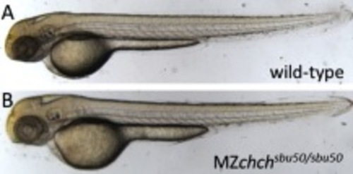
A,B: chch mutants do not show morphological abnormalities. Zebrafish embryos lacking both maternal and zygotic chch (MZchchsbu50/sbu50) do not exhibit any overt morphological defects compared with wild-type embryos at 48 hpf. PHENOTYPE:
|
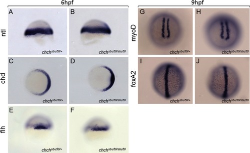
Loss of chch does not significantly alter spatial distribution of mesodermal markers during gastrulation. A–J: Whole-mount RNA in situ hybridization of early (6 hours postfertilization [hpf], A–F) and late (9 hpf, G–I) gastrula stage zebrafish embryos (A–F) using probes for mesodermal markers (as indicated). Expression of no-tail (ntl), chordin (chd), and floating-head (flh) are comparable in chch mutants and wild-type embryos at 6 hpf. Likewise expression of myoD and foxa2 (axial) are not altered chchsbu50/sbu50 mutants. Embryos were generated from a cross of a homozygous chchsbu50/sbu50 female to a heterozygous male. Genotypes of embryos were established by polymerase chain reaction following photography. |
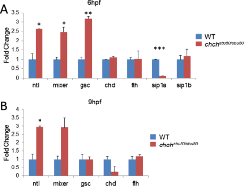
chch mutants show altered expression levels of early mesodermal and endodermal markers. Quantitative reverse transcriptase-polymerase chain reaction (qRT-PCR) analysis of transcript levels in chch mutants at 6 hours postfertilization (hpf) (gastrulation) and 9 hpf (90% epiboly). Relative comparisons were made between chchsbu50/sbu50 mutant and wild-type (WT, defined as 1) embryos. A: During blastula stage, chch mutants exhibit increased ntl, mixer, and gsc transcript levels as well as decreased sip1a expression. Levels of chd, flh, and sip1b were not significantly different between the two groups at this stage. B: chchsbu50/sbu50 embryos at 90% epiboly show increased expression of ntl and mixer while exhibiting a decrease in chd compared with wild-type. However expression of flh and gsc were unchanged. Error bars indicate standard error of the mean. Single, double, and triple asterisks indicate statistical significance (P < 0.05, P < 0.001, and P < 0.0001 respectively). EXPRESSION / LABELING:
|
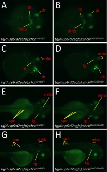
Spatial expression of the fibroblast growth factor (FGF) reporter transgenic line dusp6:d2egfp is not altered in chchsbu50/sbu50 mutant background. A-H: d2EGFP expression in siblings and chch mutants at 26 hours postfertilization (hpf) (A-D) and 50 hpf (E-H). A,B,E,F: Dorsal views showing Dusp6 reporter expression of d2EGFP. C,D,G,H: Lateral View of D2EGFP expression. Midbrain–hindbrain boundary (mhb), dorsal retina (dr), spinal neurons (spn), and trigeminal ganglion (tg) structures indicated with red text and yellow lines/brackets. |
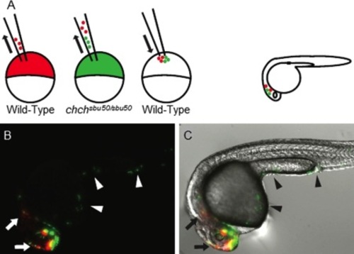
churchill mutant cells exhibit increased spreading compared with wild-type. A: A schematic of the experimental procedure. wild-type and MZchchsbu50/sbu50 donors were labeled with rhodamine (red) and fluorescein (green) dextrans, respectively, and co-transplanted into unlabeled wild-type host embryos. Host embryos were observed at 24 hpf to determine the extent of cell spreading. B,C: A fluorescent only (B) and a merge of fluorescent and DIC images (C) indicate that chch mutant cells mobilize to posterior regions of the embryo (arrowheads), whereas wild-type cells remain in or close to the head (arrows). |

Unillustrated author statements |