
- Title
-
Fss/Tbx6 is required for central dermomyotome cell fate in zebrafish
- Authors
- Windner, S.E., Bird, N.C., Patterson, S.E., Doris, R.A., and Devoto, S.H.
- Source
- Full text @ Biol. Open
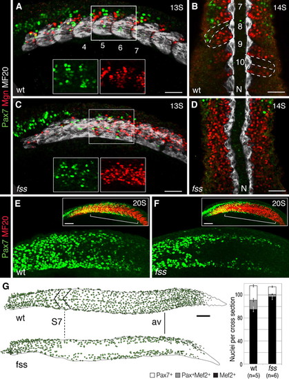
fss/tbx6 is required for the central dermomyotome development in the posterior trunk and tail. (A–D) Pax7 (green), Myogenin (Mgn, red), and MF20 (MyHC, white) expression in the anterior trunk (A,C; confocal z-stack, insets show higher magnification) and the posterior trunk (B,D) of wild type sibling (A,B) and fss/tbx6 mutant (C,D) at 13–14S stage. N, Notochordd, numbers indicate somite number. Wild type siblings express all three markers within distinct compartments of the somites (dashed lines in B). In fss/tbx6 mutants, Pax7 expressing central dermomyotome cells are restricted to the anterior trunk; the central posterior trunk shows more cells expressing Mgn. (E,F) Pax7 (green) and MF20 (MyHC, red) expression in wild type sibling (E) and fss/tbx6 mutant (F) embryo at 20S stage. Boxes show flattened confocal z-stacks of the entire trunk. In the posterior trunk Pax7 is initially expressed in cells of the dorsal and ventral dermomyotome; central Pax7 expression is delayed in wild type sibling (E), but absent in fss/tbx6 mutant (F). (G) Tracing of Pax7+ dermomyotome nuclei in representative wild type sibling and fss/tbx6 embryos at the 24h stage. In this mutant embryo, the central dermomyotome deficit/defect in fss/tbx6 mutants starts at trunk levels correlating to somite 7 in the wild type sibling (dashed line, S7). Graph showing number of Pax7+, Mef2+ and differentiating (Pax7+/Mef2+) cells (mean±s.e.m.) on sections at anal vent (av) level in wild type siblings and fss/tbx6 mutants. Scale bars: 50μm (A–D), 100μm (E–G). EXPRESSION / LABELING:
|
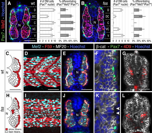
Premature dermomyotome progenitor differentiation disrupts myotome patterning. (A,B) Cross-sections and graphs showing dorsal-ventral distribution of dermomyotome (average of Pax7+ cells ± s.e.m.) and % differentiating dermomyotome (Pax7+/Mef2+) in wild type sibling and fss/tbx6 mutant (green, Pax7; red, Mef2; blue, Hoechst 33258). (C–L) Myotome morphology in wild type siblings (D–H) and fss/tbx6 (I–M) at the 24h stage. (C,H) Tracing of fast (white) and slow (red) muscle fibres on representative cross-sections. In fss/tbx6 mutants, the layer of slow fibres is disrupted and large fast muscle fibres are found lateral to the slow fibres in the central myotome. (D,E,I,J) Whole mounts (posterior trunk) and cross-sections labeled with F59 (red, labels slow myosin heavy chain at the 24h stage), Mef2 (aqua) and MF20 (white). (F,G,K,L) β-catenin labeled cross-sections double-labeled for either Pax7 (green, F,G) or 4D9 (red, engrailed, G,L); nuclei labeled with Hoechst. Arrowheads indicate large diameter, lateral fast fibres. N, notochord. Scale bars: 50 μm (A,C–E,G), 25 μm (F). Corresponding images of fss are to the same scale. |
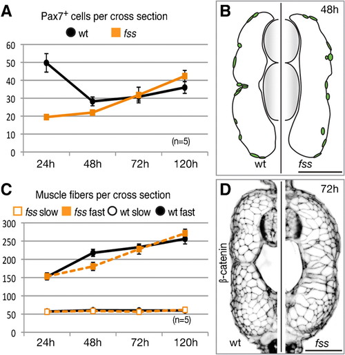
Dermomyotome recovery and muscle growth. (A) Graph showing mean number of Pax7+ cells (±s.e.m.) over time. (B) Tracing of Pax7+ dermomyotome nuclei in representative transverse section of wild type sibling (left) and fss/tbx6 mutant (right) at the 48h stage. (C) Graph showing mean number of slow and fast muscle fibres over time (± s.e.m.). (D) Cross-sections showing myotome morphology (β-catenin) at the 72h stage. Scale bars: 50 μm. PHENOTYPE:
|
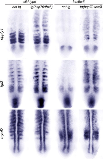
A pulse of Tbx6 expression in fss/tbx6 mutants induces segmental expression of ripply1 and fgf8, and locally represses myoD. Expression of genes expressed specifically in the anterior (ripply1, fgf8) and posterior (myoD) somite compartments in non-tg and tg(hsp70:tbx6) wild type sibling and fss/tbx6 mutant embryos. Embryos were heat shocked at the 9–10S stage, fixed at the 13–14S (myoD) and 15–16S stage (ripply1, fgf8), and flat-mounted for documentation. Scale bar: 50 μm. EXPRESSION / LABELING:
|
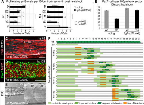
Induced Tbx6 expression differentially rescues central dermomyotome and segmentation. (A) Dorsal/ventral distribution of proliferative (phosphorylated Histone H3, pH3+) cells in non-tg and tg(hsp70:tbx6) wild type sibling and fss/tbx6 mutant embryos 8hours after heat-shocking at the 8S stage. Data are presented as mean ± s.e.m. (B) Graph showing number of Pax7+ cells in non-tg and tg(hsp70:tbx6) wild type sibling and fss/tbx6 mutant embryos at 10hours after heat-shocking at the 10S stage. (C–E) Rescue of dermomyotome and segmentation in fss/tbx6 mutant tg(hsp70:tbx6) visualized using F59 (red) and MF20 (white) (C) and Pax7 (green) and Mef2 (white) (D) labeling, and DIC imaging (E). Specimen stages are 5d (C,E) and 24h (D), rescued segments are numbered according to the corresponding region in wild-type siblings. (F) Schematic showing the location of rescued dermomyotome (light green), segments (dark green) in individual embryos after heat shocks at various times (orange). Note that a Tbx6 pulse rescues the formation of 1–3 somite boundaries and central dermomyotome over 8–10 somite lengths. Scale bars: 100μm (C,D), 50μm (E). PHENOTYPE:
|