
- Title
-
SUMO1-activating enzyme subunit 1 is essential for the survival of hematopoietic stem/progenitor cells in zebrafish
- Authors
- Li, X., Lan, Y., Xu, J., Zhang, W., and Wen, Z.
- Source
- Full text @ Development
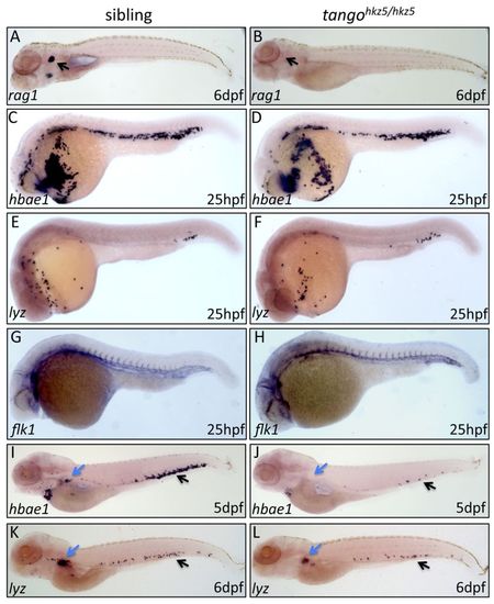
Impairment of definitive hematopoiesis in tangohkz5 mutant zebrafish. (A,B) WISH of rag1 in 6 dpf tangohkz5/hkz5 embryos and wild-type siblings. Arrows indicate the thymus. (C-H) WISH of hbae1, lyz and flk1 in 25 hpf tangohkz5/hkz5 mutant embryos and wild-type siblings. (I-L) WISH of hbae1 and lyz in 5 dpf and 6 dpf tangohkz5/hkz5 mutant embryos and wild-type siblings. Blue arrows indicate the position of the kidney; black arrows indicate the CHT. |
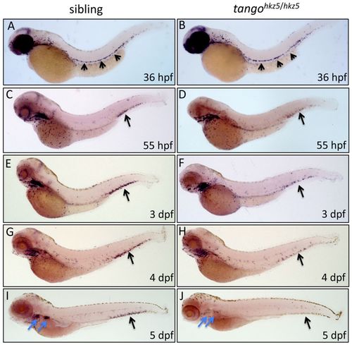
HSPCs are depleted in the CHT of tangohkz5 mutant zebrafish. (A-J) WISH of cmyb in homozygous mutants of tangohkz5 and wild-type siblings at 36 hpf (A,B), 55 hpf (C,D) and 3-5 dpf (E,J). Arrows in A and B indicate the AGM region. Black arrows in C-J represent the CHT. The anterior and posterior blue arrows in I-J indicate the thymus and kidney, respectively. |
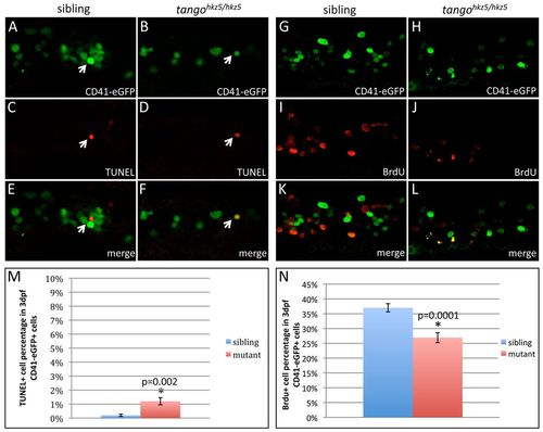
tangohkz5 mutation causes an increased apoptosis and a decreased proliferation of HSPCs. (A-F) Double immunostaining of CD41-eGFP and TUNEL reveals a slight increase of apoptosis of CD41-eGFP+ cells in the CHT of 3 dpf tangohkz5/hkz5 mutant zebrafish embryos. Arrows show the CD41-eGFP+ and TUNEL+ cells. (G-L) Double immunostaining of CD41-eGFP and BrdU shows a reduced proliferation of CD41-eGFP+ cells in the CHT of 3 dpf tangohkz5/hkz5 mutant embryos. (M) Quantification of the percentage of TUNEL+ cells in the 3 dpf CD41-eGFP+ cell populations. (N) Quantification of the percentage of BrdU+ cells in the 3 dpf CD41-eGFP+ population. Error bars represent s.e.m. PHENOTYPE:
|
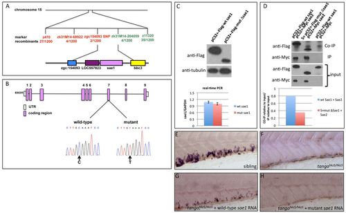
The tangohkz5 mutant phenotype is caused by a nonsense mutation in sae1. (A) The tangohkz5 mutation was mapped on chromosome 15 between the SNP marker zgc154093 (two recombinants out of 1200 tangohkz5/hkz5 embryos) and zk31M14-204059 (four recombinants out of 1200 tangohkz5/hkz5 embryos). This region contains four genes: sae1, LOC557823, bbc3 and zgc154093. (B) The sae1 gene contains nine exons and a C-to-T substitution was found in exon 7. (C) Western blotting of 293T cells transfected with pCS2+-Flag-wt sae1 or pCS2+-Flag-mut Δsae1. The expression level of the mutant ΔSae1 protein was much lower than that of wild-type Sae1. Tubulin was used as a loading control. Real-time PCR showed the similar RNA levels of wild-type and mutant forms of sae1 relative to Gapdh (P=0.3, n=3). (D) Co-immunoprecipitation assay. 293T cells were co-transfected with either pCS2+-Flag-wt sae1/pCS2+-Myc-sae2, or fivefold pCS2+-Flag-mut Δsae1/pCS2+-Myc-sae2. Transfection with pCS2+-Flag-wt sae1/pCS2+-Myc was used as a negative control. The input showed the similar expression levels of either Flag-Sae1 or Myc-Sae2 between wild type and mutant. Quantification showed that the co-immunoprecipitated mutant ΔSae1 was decreased by >50% compared with wild-type Sae1. The data were reproducible in two independent experiments. (E-H) WISH shows the cmyb expression in the CHT of wild-type siblings (E), tangohkz5/hkz5 embryos (F), tangohkz5/hkz5 embryos injected with sae1 wild-type mRNA (G), and tangohkz5/hkz5 embryos injected with the mutant Δsae1 mRNA (H). Error bars represent s.e.m. |
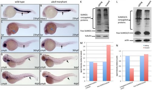
Loss of Sae1 function impairs the sumoylation pathway. (A-D) WISH shows the hbae1 and lyz expression in 23 hpf wild-type fish and ubc9 morphants. Black arrows indicate the ICM. Blue arrows indicate the yolk sac. (E,F) WISH indicates the cmyb expression in 36 hpf wild-type fish and ubc9 morphants. Arrows indicate the AGM of zebrafish. (G-J) WISH shows the cmyb expression in 3-4 dpf wild-type fish and ubc9 morphants. Arrows indicate the CHT region. (K) Western blotting indicates the expression of free SUMO1s and the SUMO1 conjugates in 6 dpf siblings (left) and tangohkz5/hkz5 mutant embryos (right). (L) Western blotting shows the expression of free SUMO2/3 and the SUMO2/3 conjugates in 6 dpf siblings (left) and tangohkz5/hkz5 mutant embryos (right). (M) Quantification of the SUMO1 conjugates and the free SUMO1 relative to tubulin. (N) Quantification of the SUMO2/3 conjugates and the free SUMO2/3 relative to actin. EXPRESSION / LABELING:
PHENOTYPE:
|
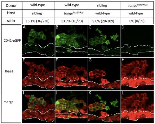
Sae1 cell-autonomously regulates HSPC survival in the CHT. (A-D) Immunofluorescence staining of GFP in the CHT of 5 dpf host fish. (E-H) Immunofluorescence staining of Hbae1 in the CHT of 5 dpf host fish. (I-L) Merged images of GFP and Hbae1 immunofluorescence staining. A, E and I are the tangohkz5 sibling hosts transplanted with wild-type cells carrying CD41-eGFP. B, F and J are the tangohkz5/hkz5 mutants transplanted with wild-type cells carrying CD41-eGFP. C, G and K are the wild-type fish transplanted with tangohkz5 sibling cells carrying CD41-eGFP. D, H and L are the wild-type fish transplanted with tangohkz5/hkz5 mutant cells carrying CD41-eGFP. White dotted lines indicate the caudal vein. |
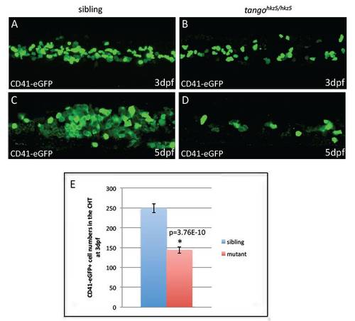
CD41-eGFP+ HSPCs are reduced in the CHT of tangohkz5 mutants. (A-D) Immunostaining of eGFP in the CHT of tangohkz5/hkz5 mutant embryos and their siblings at 3 dpf and 5 dpf. (E) Quantification of CD41eGFP+ cell number in the CHT of 3 dpf tangohkz5/hkz5 mutant embryos and siblings. Error bars represent s.e.m. PHENOTYPE:
|
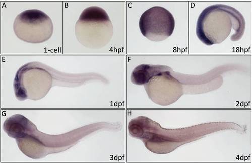
The expression pattern of sae1 during zebrafish early development. (A-H) WISH shows sae1 expression in wild-type zebrafish at the 1-cell stage (A), 4 hpf (B), 8 hpf (C), 18 hpf (D), 1 dpf (E), 2 dpf (F), 3dpf (G) and 4 dpf (H). |
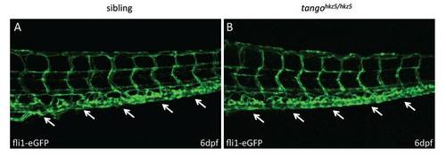
The CHT vascular plexuses are intact in tangohkz5 mutants. (A,B) Anti-GFP staining reveals a similar overall structure of the CHT vascular plexuses (arrows) in the 6 dpf Tg(fli1-eGFP)tangohkz5/hkz5 mutants (B) and their siblings (A). |

Unillustrated author statements |