
- Title
-
In vivo conditions to identify prkci phosphorylation targets using the analog-sensitive kinase method in zebrafish
- Authors
- Cibrián Uhalte, E., Kirchner, M., Hellwig, N., Allen, J.J., Donat, S., Shokat, K.M., Selbach, M., and Abdelilah-Seyfried, S.
- Source
- Full text @ PLoS One
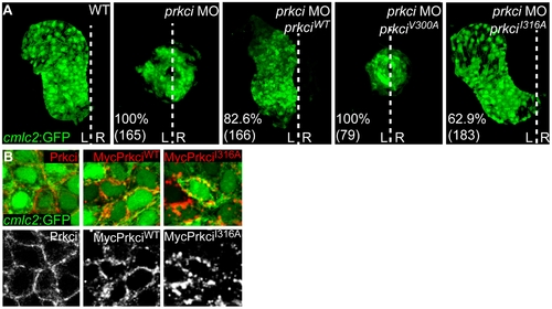
Mutant PrkciI316A has normal in vivo biological activity. (A) Reconstruction of confocal Z-stack sections of embryonic hearts at 28–30 hpf. Transgenic Tg[cmlc2:GFP]twu34 one-cell stage embryos were injected with prkci MO alone or together with mRNA encoding PrkciWT or analog-sensitive mutant forms of Prkci. Whereas the wild-type heart elongates into a heart tube and towards the left during cardiac jogging, heart development arrests at the cone stage and the heart remains at the embryonic midline in prkci morphants. In functional rescue experiments, injection of prkciMO together with mRNA encoding HisMyc-PrkciWT or PrkciI316A rescues heart tube elongation. In comparison, the analog-sensitive mutant form PrkciV300A fails to rescue heart tube formation in prkci morphants. Percentiles indicate the occurrence of the most common phenotype as depicted in the images and numbers show the total of embryos tested. White dotted line indicates the embryonic midline. L, left; R, right. (B) Membrane localization of endogenous Prkci detected with an anti-Prkci antibody and exogenous PrkciWT or PrkciI316A in zebrafish cardiomyocytes detected with an anti-Myc antibody. Images are confocal reconstructions of single Z-stack sections of embryonic hearts marked by the transgenic reporter Tg[cmlc2:GFP]twu34 at 28–30 hpf. Expression of exogenous HisMyc-PrkciWT or HisMyc-PrkciI316A in cardiomyocytes reveals that both recombinant proteins localize to the cell membrane. |
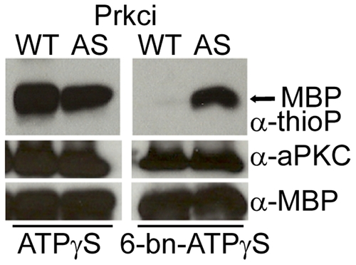
Mutant PrkciI316A uses bulky N6-benzyl ATPγS. Kinase reaction with Myelin Basic Protein (MBP) and ATPγS or N6-benzyl ATPγS (6-bn- ATPγS), followed by PNBM alkylation. In comparison, only mutant PrkciI316A efficiently utilizes N6-benzyl ATPγS to thiophosphorylate MBP. Labeled MBP is detected by Western blot analysis with rabbit monoclonal anti-thiophosphoester antibody (α-thioP). |
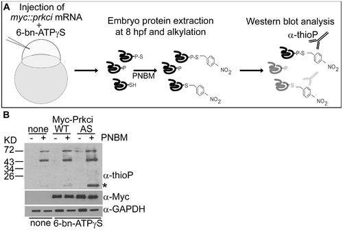
Thiophosphorylation of substrate proteins by PrkciI316 in the zebrafish embryo. (A) Schematic diagram of the in vivo labeling method for the selective labeling of PrkciI316A substrates during zebrafish development. (B) In vivo thiophosphorylation in zebrafish embryos injected at the one-cell stage with 200 μM N6-benzyl-ATPγS (6-bn-ATPγS) and mRNA encoding either PrkciWT or PrkciI316A (AS). Western blot analysis with rabbit monoclonal anti-thiophosphoester (α-thioP) C51-8 antibody (Epitomics) of 80% epiboly (6–8hpf) samples alkylated with 2.5 mM PNBM reveals a selectively labeled protein in the PrkciI316A (AS) sample (asterisk). |