
- Title
-
Expression analysis of zebrafish membrane type-2 matrix metalloproteinases during embryonic development
- Authors
- Quick, R.E., Dunlap, J.A., and Jessen, J.R.
- Source
- Full text @ Gene Expr. Patterns
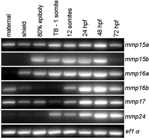
mRNA expression analysis of zebrafish membrane tethered mmps. RT-PCR was used to amplify mmp cDNA sequences generated from 1 µg total RNA collected at multiple embryonic stages. PCR was performed for 35 cycles because 40 cycles amplified saturating amounts of DNA product. Equal volumes of PCR product were loaded onto an agarose gel and detected using ethidium bromide and UV transillumination. To detect maternal mmp gene expression, total RNA was isolated from <8-cell stage embryos. hpf, hours post-fertilization; TB, tailbud. EXPRESSION / LABELING:
|
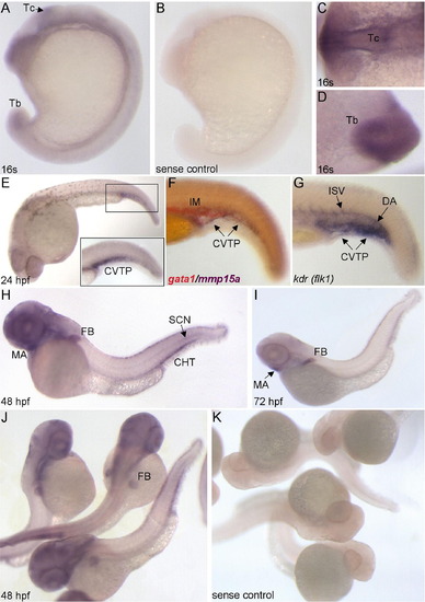
Expression pattern of mmp15a examined using whole-mount RNA in situ hybridization. (A and B) Lateral views of 16-somite stage (16s) embryos with ventral to the left and anterior to the top. Close-up dorsal view of the tectum, Tc (C) and ventral view of the tailbud, Tb (D). (E–G) Lateral views of 24 hpf embryos labeled for mmp15a, gata1/mmp15a, and kdrl (flk1). Inset in panel (E) shows close-up of the cardinal vein tail plexus, CVTP. (H, J, and K) Lateral/dorsal views of 48 hpf embryos with anterior to the left (H). (I) Lateral view of 72 hpf embryo with anterior to the left. CHT, caudal hematopoietic tissue; DA, dorsal aorta; FB, fin bud; IM, intermediate cell mass; ISV, intersegmental vessels; MA, mandibular arch; SCN, spinal cord neurons. |
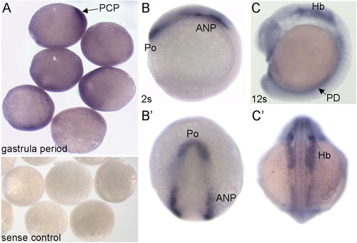
Expression pattern of mmp15b during early development. (A) Gastrula period embryos showing ubiquitous mmp15b expression with stronger staining in the prechordal plate mesendoderm (PCP). Gastrula-stage embryos labeled with mmp15b sense control probe are shown at the bottom. Lateral (B and C) and dorsal (B′ and C′) views of 2-somite (2s) and 12-somite (12s) stage embryos. ANP, anterior neural plate; Hb, hindbrain; Po, polster; PD, pronephric duct. EXPRESSION / LABELING:
|
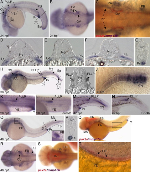
Expression pattern of mmp15b during pharyngula and hatching periods. (A) Lateral view of 24 hpf embryo with anterior to the left. Black lines denote cross-section slices shown in panels (D–G). (B and C) Dorsal views of 24 hpf embryos with anterior to the left. Embryo in panel (C) is double-labeled with mmp15b and pax2a (red). (D–G) Cross-sections of 24 hpf embryos at positions marked in panel (A). (H and J) Lateral views of 36 hpf embryos with anterior to the left. (I) Cross-section of 36 hpf embryo trunk (position shown in panel H) showing expression in posterior lateral line primordia, PLLP. Mmp15b and cxcr4b expression at 28 hpf (K and L) and 42 hpf (M and N), lateral views with anterior to the left and dorsal to the top. YE, yolk extension. (O, Q, and T) Lateral views of 48 hpf embryos with anterior to the left. Inset in panel (O) shows close-up of mmp15b expression in the fin bud, FB. (P) Cross-section through the proctodeum (Pr) of a 48 hpf embryo (position shown in panel O). (R and S) Dorsal views of 48 hpf embryos with anterior to the left. (Q, S, and T) Double labeling with pax2a (red) highlights mmp15b expression in pharyngeal arches (PA) and pronephric duct, PD. CN, caudal notochord; Ep, epidermis; Hb, hindbrain; MA, mandibular arch; My, myoseptum; Nc, notochord; OV, otic vesicle; PP, pharyngeal pouch. |
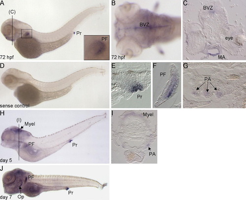
Expression pattern of mmp15b in larval stage embryos. (A and D) Lateral and dorsal (B) views of 72 hpf embryos with anterior to the left. Inset in panel (A) shows close-up of pectoral fin, PF. (C) Cross-section through the brain ventricular zone (BVZ) and mandibular arch (MA) of a 72 hpf embryo. (E–G) Sagittal plane sections of 72 hpf embryos showing mmp15b expression in the proctodeum (Pr), PF, and anterior pharyngeal arches, PA. (H and J) Lateral views of day 5 and 7 embryos with anterior to the left. (I) Cross-section through the myelencephalon (Myel) and PA of a day 5 embryo (position shown in panel H). Op, operculum. EXPRESSION / LABELING:
|
Reprinted from Gene expression patterns : GEP, 12(7-8), Quick, R.E., Dunlap, J.A., and Jessen, J.R., Expression analysis of zebrafish membrane type-2 matrix metalloproteinases during embryonic development, 254-260, Copyright (2012) with permission from Elsevier. Full text @ Gene Expr. Patterns