
- Title
-
The zebrafish sf3b1(b460) mutant reveals differential requirements for the sf3b1 pre-mRNA processing gene during neural crest development
- Authors
- An, M., and Henion, P.D.
- Source
- Full text @ Int. J. Dev. Biol.
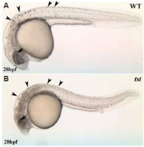
Visible live phenotype of tstb460 mutants. Neural crest-derived melanophores; see arrowheads in (A) are absent in tstb460 mutants; see arrowheads in (B). In contrast, melanized pigmented retinal epithelial cells are present in the mutants (arrow). PHENOTYPE:
|
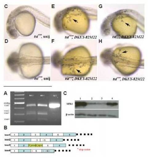
Molecular consequences of sf3b1 mutations (A,B).Variant transcripts of sf3b1 in tstb460 mutants. (A) RT-PCR products were amplified between exon1 and exon6 of sf3b1 with 20-24 hpf AB wildtype and tstb460 homozygous mutant total RNA as template using Sf3b1aa (46)F and Sf3b1aa (579)R primers. The single AB wildtype RT-PCR product was 534 bp (tran1). tstb460 homozygous mutants contained 4 different transcripts; tran1(534 bp), and variant transcripts tran2(412 bp), tran3(390 bp), and tran4(312 bp). (B) Diagram of variant transcripts in tstb460 homozygous mutants. (C) Western blot analysis of Sf3b1 protein levels in wildtype, tstb460 mutant and hi3394a mutant embryos at 24 hpf. Lane 1: wildtype embryos; Lane 2: tstb460 homozygous mutants; Lane 3: hi3394a homozygous mutants; Lane 4: hsp>sf3b1 injected tstb460 mutants with melanophore phenotype rescue. EXPRESSION / LABELING:
PHENOTYPE:
|
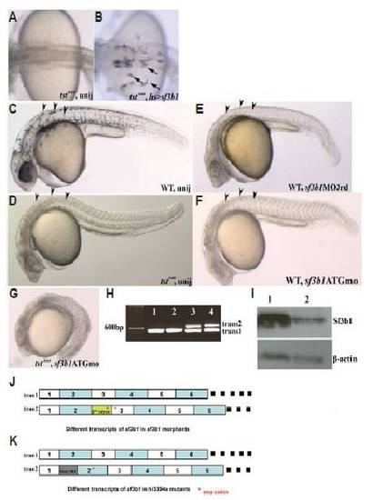
sf3b1 gain- and loss-of-function analyses. (A,B) The tstb460 phenotype is partially rescued by sf3b1 misexpression. (A) Melanophores are absent in uninjected tstb460 mutant embryos. (B) tstb460 homozygous mutant embryos were injected with hs>sf3b1 and heat-shocked 2 or 3 times (6 hpf, 10 or 11 hpf, 22 or 23 hpf), 1h/each time. Several melanophores present in the dorsal trunk region and over the yolk region in injected tstb460 mutant embryos (arrows). (C-F) Phenocopy of tstb460 mutant embryos using morpholino knockdown analysis. (C) Lateral view of a live uninjected wildtype embryo at 30 hpf. (D) Lateral view of a tstb460 homozygous mutant at 30 hpf. (E) Lateral view of a live sf3b1MO3rd morphant showing the absence of melanophores (arrowheads) and CNS (brain) cell death at 30 hpf. (F) Lateral view of a live sf3b1MOATG morphant showing phenocopy of the tstb460 mutant at 30 hpf. (G) The phenotype was more severe in the sf3b1MOATG injected tstb460 mutant as compared with uninjected tstb460 mutant, suggesting that the tstb460 allele is hypomorphic. (H) Variant transcripts in sf3b1MO3rd morphants. Lane1 and lane2: RT-PCR product from uninjected wildtype total RNA. Lane 3 and lane 4: RT-PCR product from sf3b1MO3rd morphant total RNA. (I) Western blot analysis of Sf3b1 protein levels in uninjected wildtype embryos and sf3b1MOATG morphants at 12 hpf. Lane 1: uninjected wildtype embryos; Lane 2: sf3b1MOATG morphants. (J) Schematic diagram of different sf3b1 transcripts in sf3b1MO3rd morphants. (K) Schematic diagram of different sf3b1 transcripts in hi3394a mutants. PHENOTYPE:
|
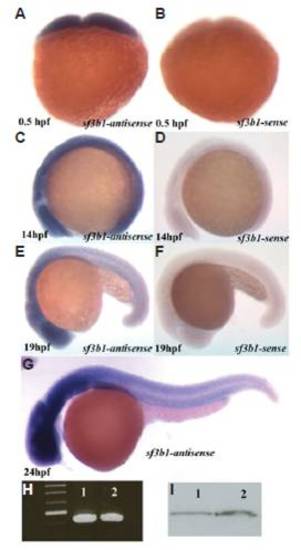
Whole-mount in situ hybridization with sf3b1 antisense probe and sense probes in wildtype embryos. (A,C) sf3b1 is ubiquitously expressed at 0.5 hpf and 14 hpf. (E,G) The expression of sf3b1 appears elevated in the brain, neural tube and ventral trunk region at 19 hpf and 24 hpf (lateral view, anterior to left). (B,D,F) sf3b1 sense controls. (H) RT-PCR products using total RNA from 0.5hpf (lane1) and 24hpf (lane2) wildtype embryos as templates and Sf3b1aa(46)F and Sf3b1aa(579)R as primers. (I) Western blot analysis shows Sf3b1 protein present maternally. Lane1: protein from 24 hpf wildtype embryos as control. Lane2: protein from 1 hpf wildtype embryos. |
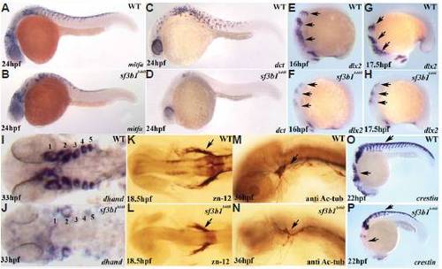
Neural crest progenitor and cranial ganglia phenotypes of sf3b1b460 mutants. There is reduced melanoblast expression of mitfa (A,B) and nearly absent melanoblast expression of dct (C,D) in sf3b1b460 mutants. (E-J) Pharyngeal arches are dramatically reduced in sf3b1b460 mutant embryos. (E-H) Lateral view of whole-mount in situ embryos with dlx2 antisense RNA probe, arch progenitors denoted by arrows. (I,J) Dorsal view of flat-mounted in situ embryos with dhand antisense RNA probe, pharyngeal arches 1 and 2 are decreased and 3, 4 and 5 are absent in sf3b1b460 mutants. (K-N) the cranial ganglia are also affected in sf3b1b460 mutants. (K,L) Whole-mount antibody staining with zn-12. Arrows in (K,L) show the trigeminal ganglia. (M,N) Whole-mount antibody staining with anti-acetylated tubulin antibody. Arrows in (M,N) show the decreased and disorganized trigeminal ganglia in sf3b1b460 mutants compared to wildtype siblings. (O,P) Crestin expressing cells are present in sf3b1b460 mutant embryos. However, these cells are reduced in numbers, particularly in the cranial region and fail to migrate normally in the sf3b1b460 mutant (P) compared to wildtype embryos (O). EXPRESSION / LABELING:
PHENOTYPE:
|
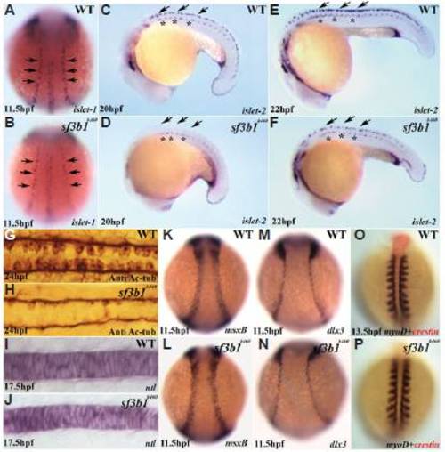
Early development of ectodermal and mesodermal derivatives in sf3b1b460 mutants. (A,B) Whole-mount in situ hybridization with islet-1 antisense RNA probes shows that the number of Rohon-Beard precursor cells (arrows) is decreased in sf3b1b460mutants (dorsal view, anterior to top). (C-F) Whole-mount in situ hybridization with islet-2 antisense RNA probe shows deficiency in the development of Rohon-Beard sensory neurons (arrows), whereas the development of primary motor neurons is comparatively less affected in mutant embryos (asterisks). (G,H) Dorsal view of spinal cords labeled with anti-acetylated tubulin (Anti Ac-tub), anterior to left, showing that the Rohon-Beard sensory neuron phenotype persists at later stages in sf3b1b460 mutants. K-N: Induction of the neural plate border occurs normally in sf3b1b460 mutants. Whole-mount in situ hybridization with msxB (K,L) and dlx3 (M,N). (I,J;O,P) The derivatives from paraxial mesoderm and chordamesoderm are not affected in sf3b1b460mutants. (I,J) Lateral view of whole-mount in situ with ntl, anterior to left. (O,P) Dorsal view of double in situ with crestin (red) and myoD (blue), anterior to top. EXPRESSION / LABELING:
PHENOTYPE:
|
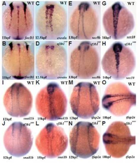
Significant abnormalities in trunk neural crest expression of critical transcriptional regulators as well as crestin at early developmental stages in sf3b1b460 mutants, but grossly normal expression in the cranial neural crest. Whole-mount in situ hybridization with foxd3 (A,B), crestin (C,D), sox9b (E,F), sox10 (G,H), snai1b (I-L), and tfap2a (M-P) antisense RNA probes. All embryos are dorsal view, anterior to top with the exception of (O,P) which is anterior to left. EXPRESSION / LABELING:
|
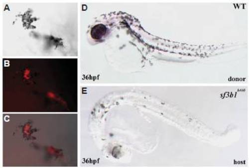
Genetic mosaics of sf3b1b460 mutants and wildtype embryos. (A) Melanophores derived from wildtype donor cells on the yolk of a sf3b1b460 mutant host (DIC image). (B) Fluorescence image of the same (donor-derived) cells. (C) (A,B) merged. (D) A wildtype donor. (E) An sf3b1b460 mutant host with wildtype donor-derived melanophores. |
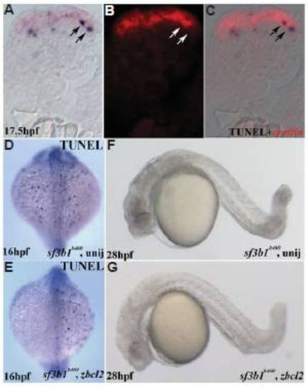
Analysis of cell death in sf3b1b460 mutants. (A-C) Double labeling with TUNEL (NBT/BCIP) and crestin whole-mount in situ hybridization (fast red). Transverse section is from the trunk region of a sf3b1b460 mutant, dorsal is up. (A) DIC image/TUNEL; (B) fluorescence image/crestin of the same section; (C) (A,B) merged. (D) Dorsal view of uninjected sf3b1b460 mutant at 16 hpf with TUNEL staining. (E) Dorsal view of zbcl2 mRNA injected sf3b1b460 mutant at 16 hpf with TUNEL staining. Reduced TUNEL staining is evident on the left side. (F) Lateral view of live uninjected sf3b1b460 mutant at 28 hpf. (G) Lateral view of live zbcl2 injected sf3b1b460 mutant at 28 hpf, showing less CNS cell death as compared with uninjected sf3b1b460 mutants but no melanophore rescue. PHENOTYPE:
|
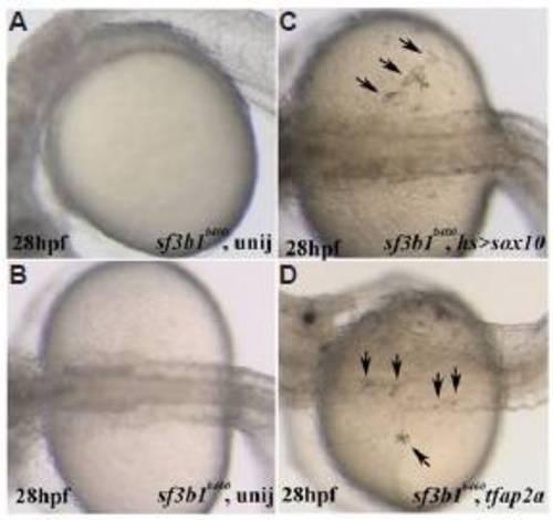
Misexpression of genes that regulate neural crest development results in melanophore rescue in sf3b1b460 mutant embryos. (A) Lateral view of live uninjected sf3b1b460 mutant at 28 hpf (anterior to left). (B) PHENOTYPE:
|