Genetic suppressor screens in zebrafish reveal additional mutations capable of rescue. (a) The bacterial artificial chromosome (BAC) transgenic construct containing a wild-type Tif1gamma locus and green fluorescent protein (GFP) driven by an actin promoter (Pactin) used in our recent genetic suppressor screen [2]. The transgene was injected into one-cell-stage embryos (right) to rescue the lethality of Tif1gamma mutant (mon) fish. (b) Schematic diagram of the suppressor screen. Stable transgenic fish are homozygous mutants for the endogenous tif1gamma locus (mon/mon) but retain viability because they are heterozygous for the transgene. The GFP marker on the transgene makes them green fluorescent. F0 males were mutagenized with ethylnitrosourea (ENU). In the F1 generation, 25% of progeny were transgene homozygotes (Tg homo, mon/mon; Tg/Tg, bright green), 50% were transgene heterozygotes (Tg het, mon/mon; Tg/+, light green, in red circle), and 25% lacked the transgene (No Tg, mon/mon, gray). Only the progeny that were heterozygous for the transgene were raised to adults. The F1 females were then squeezed to provide unfertilized eggs that were activated by UV-treated sperm. The UV treatment destroys the paternal DNA while still allowing fertilization. The resulting F2 embryos were haploid and were subjected to in situ hybridization (ISH) at 22 hours post-fertilization for GFP and beta e3 globin probes. Transgenic embryos (mon;Tg) were positive for both probes, whereas non-transgenic embryos (mon) were negative for both probes. However, embryos that were negative for GFP but positive for globin indicated the presence of a genomic suppressor (sup) mutation. Modified, with permission, from [2].
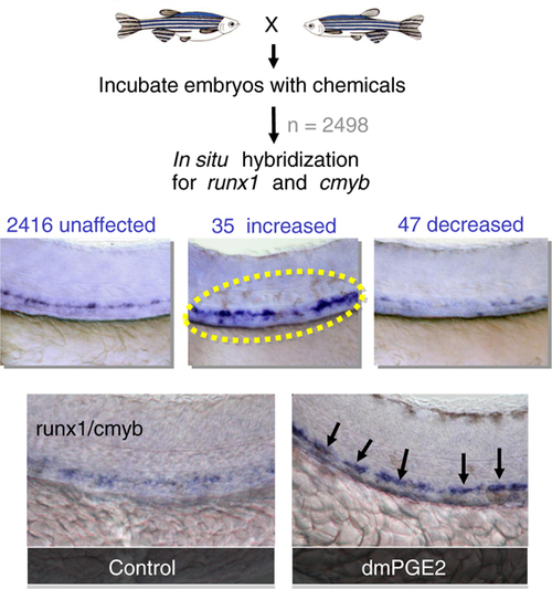
Large-scale vertebrate chemical screening made possible by zebrafish. Embryos are incubated in groups of 5-10 with approximately 2,500 different chemicals. At 36 hours post-fertilization, in situ hybridization is conducted to analyze the expression of early hematopoietic markers such as runx1 and c-myb. The embryos are then scored for a change in hematopoietic expression. We recently used this technique [45] to identify 82 compounds that influence hematopoietic stem cell differentiation, the most prominent of which was dimethyl prostaglandin E2 (dmPGE2). Modified with permission, from [45].
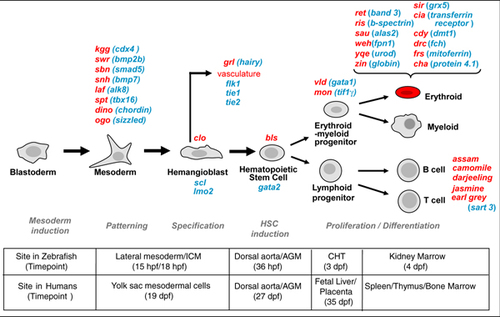
Hematopoiesis in zebrafish and humans, and known zebrafish blood mutants. The stages of hematopoiesis are illustrated, with the genes and mutants identified as affecting each stage shown (red, zebrafish blood mutants; blue, genes altered by the mutations) and the processes in bold below. Bottom: the sites and times of the events shown in human and zebrafish. AGM, aorta gonad mesonephros; CHT, caudal hematopoietic tissue; dpf, days post-fertilization; hpf, hours post-fertilization; HSC, hematopoietic stem cell.
Acknowledgments
This image is the copyrighted work of the attributed author or publisher, and
ZFIN has permission only to display this image to its users.
Additional permissions should be obtained from the applicable author or publisher of the image.
Full text @ Genome Med.
Your Input Welcome
Thank you for submitting comments. Your input has been emailed to ZFIN curators who may contact you if
additional information is required.
Oops. Something went wrong. Please try again later.