
- Title
-
Combinatorial roles for BMPs and Endothelin 1 in patterning the dorsal-ventral axis of the craniofacial skeleton
- Authors
- Alexander, C., Zuniga, E., Blitz, I.L., Wada, N., Le Pabic, P., Javidan, Y., Zhang, T., Cho, K.W., Crump, J.G., and Schilling, T.F.
- Source
- Full text @ Development
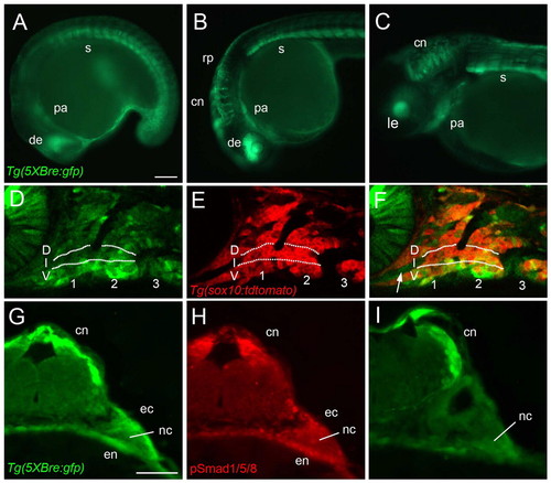
Expression of Tg(Bre:GFP) in the pharyngeal arches. (A-C) GFP fluorescence in living Tg(Bre:GFP) transgenic embryos, lateral views, anterior towards the left. (D-F) Lateral views of the arches at higher magnification. Confocal slices of Tg(Bre:GFP) (D) and Tg(sox10:lyn-tdTomato) (E) double transgenics. The two channels are merged in F, demonstrating direct BMP responses in NC and in the stomodeum (arrow). (G-I) Adjacent transverse sections through the hindbrain and arches stained with anti-GFP (G,I) and anti-pSmad1/5/8 (H), revealing BMP responses in the NC and in surrounding endoderm (en) and ectoderm (ec). (A) 16 hpf. (B) 24 hpf. (C) 48 hpf. (D-F) 28 hpf. (G-I) 30 hpf. cn, commissural neurons; D, dorsal; de, dorsal eye; ec, ectoderm; en, endoderm; I, intermediate; le, lens; nc, neural crest; pa, pharyngeal arches; rp, roofplate; s, somites; V, ventral. Scale bars: 100 μm. |
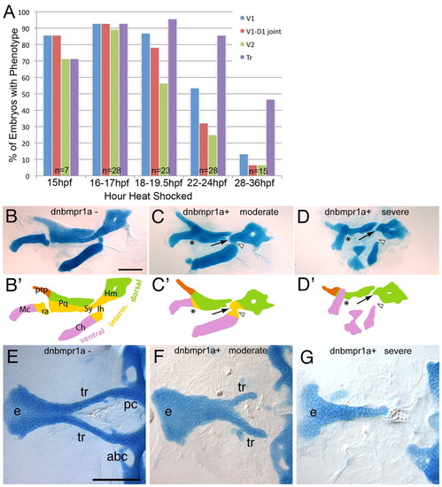
Requirements for BMP signaling in pharyngeal cartilage and palate development. (A) Histogram quantifying the frequency of defects in different skeletal elements caused by heat shocking Tg(hs:dnBmpra1-GFP) embryos at different stages between 15 and 36 hpf. (B-G) Alcian Blue stained cartilages of 5 dpf larvae, dissected and flat-mounted, anterior towards the left. (B-D) Pharyngeal cartilages of control (B), moderate (C) and severely affected (D) Tg(hs:dnBmpra1-GFP) transgenics, heat shocked at 16-18 hpf, lateral views. (B2-D2) Diagrams of cartilage elements in arch 1 (mandibular, Mc and Pq) and arch 2 (hyoid, Ch, Ih and Hm) corresponding to cartilages in B-D. Lateral views, anterior towards the left. Colors indicate dorsal (green), intermediate (yellow) and ventral (pink) elements. Arrows indicate dorsal arch 2 elements; asterisks indicate fused joints in arch 1; arrowheads indicate joints in arch 2. (E-G) Neurocranial cartilages of the palate, ventral view: control (E), moderate (F) and severe (G). abc, anterior basicranial commissure; Ch, ceratohyal; e, ethmoid plate; Hm, hyomandibular; Ih, interhyal; Mc, Meckels cartilage; pc, parachordals; Pq, palatoquadrate; ptp, pterygoid process; ra, retroarticular process of Meckel’s cartilage; Sy, symplectic; tr, trabeculae. Scale bars: 100 μm. |
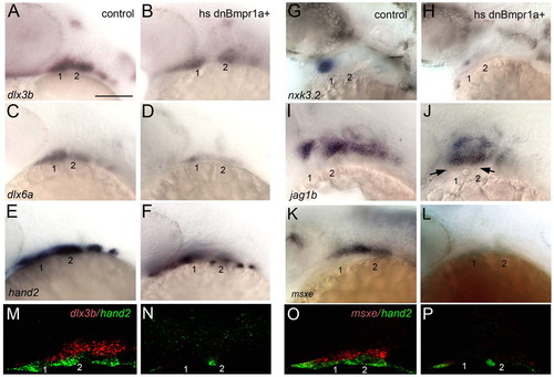
BMP signaling is required for ventral cell specification in the arches. (A-P) Whole-mount in situ hybridization to detect expression of genes involved in DV arch patterning, lateral views, anterior towards the left in controls (A,C,E,G,I,K,M,O) and Tg(hsp70I:dnBmpra1-GFP) transgenics heat shocked at 16-18 hpf (B,D,F,H,J,L,N,P). dlx3b (A,B), dlx6a (C,D) and hand2 (E,F) expression is reduced at 28 hpf. nkx3.2 (G,H) expression is reduced and jag1b expression (I,J) expands ventrally at 36 hpf (arrows). msxe expression is lost at 30 hpf (K,L). Arches 1 and 2 are numbered. (M-P) Two-color fluorescent in situs to detect dlx3b (M,N, red) or msxe (O,P, red) simultaneously with hand2 (green). Scale bar: 100 μm. |
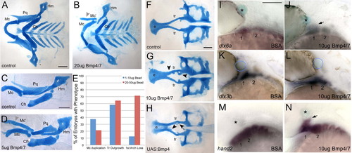
Exogenous BMP is sufficient to induce ventral arch identity. (A-D,F-H) Alcian Blue-stained cartilages of 4-5 dpf larvae, dissected and flat-mounted, anterior towards the left. (I-N) Whole-mount in situ hybridization at 30 hpf, lateral views, anterior towards the left. (A,B) Pharyngeal cartilages of a control (A) and an embryo implanted behind the eye at 20 hpf with a bead soaked in 20 μg/μl of human recombinant BMP4/7 heterodimers (B). (C,D) Isolated cartilages of the mandibular (1) and hyoid (2) arches: control (C); BMP4/7 bead-implanted (D). Grey arrowheads in B,D indicate duplicated Mc cartilages. (E) Histogram of phenotype frequency in bead-implanted embryos depending on BMP4/7 concentration. (F-H) Neurocranial cartilages: control (F); BMP4/7 bead-implanted (G); heat-shocked Tg(hsp70I:Gal4;UAS:Bmp4) (H). Black arrowheads indicate ectopic cartilages. (I,J) dlx6a expression slightly expands dorsally (arrow) in response to a BMP4/7-soaked bead (J). (K,L) dlx3b expression does not change when BMP4/7-soaked beads are implanted. (M,N) hand2 expression also expands dorsally (arrow) in response to BMP4/7 protein. Asterisks and blue circles indicate beads. 1, mandibular arch; 2, hyoid arch; Ch, ceratohyal; e, ethmoid plate; Hm, hyomandibular; Mc, Meckel’s cartilage; Mc’, ectopic Meckel’s cartilage; Pq, palatoquadrate; tr, trabeculae. Scale bars: 100 μm. |
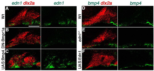
BMPs regulate Edn1 expression. Projections of double fluorescent in situ hybridization, lateral views, anterior towards the left. (A-C) dlx2a (red) and edn1 (green) in wild-type (A), heat shocked Tg(hsp70I:dnBmpr1a-GFP) (B) and heat shocked Tg(hsp70I:Gal4;UAS:Bmp4) (C) embryos. (D-F) dlx2a (red) and bmp4 (green) in wild-type (D), edn1–/– mutant (E) and heat shocked Tg(hsp70I:Gal4;UAS:Edn1) (F) embryos. Scale bar: 50 μm. |
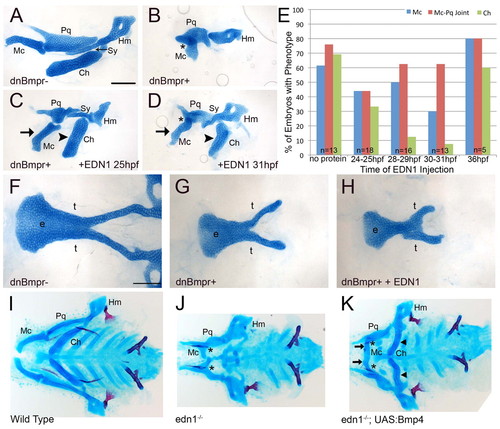
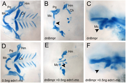
Exogenous Edn1 rescues BMP deficiency and vice versa. (A-D,F-K) Alcian Blue stained cartilages of 5 dpf larvae, anterior towards the left. (A-D) Dissected cartilages, lateral views, of a control (A), Tg(hsp70I:dnBmpr1a-GFP) transgenic heat shocked at 16-18 hpf (B) and similarly treated transgenics in which 0.5 μg human recombinant EDN1 protein was microinjected into the arches at 25 hpf (C) and 31 hpf (D). (E) Histogram depicting percentages of Tg(hs:dnBmpra1-GFP)+ embryos with severe ventral arch defects without and with EDN1 protein injected. (F-H) Ventral view of neurocranial cartilage in control (F), dnBmpr+ (G) and dnBmpr+ with 0.5 μg EDN1 injected (H). (I-K) Whole-mount cartilages, ventral views, of non-transgenic wild-type control (I), non-transgenic edn1–/– mutant (J) and edn1–/–; Tg(hsp70I:Gal4;UAS:Bmp4) (K) embryos subjected to a 1-minute heat shock at 21 hpf. Arrows show rescued ventral Mc; arrowheads show rescued ventral Ch cartilages; asterisks indicate fused Mc-Pq joints. Ch, ceratohyal; e, ethmoid plate; Hm, hyomandibular; Mc, Meckels cartilage; Pq, palatoquadrate; Sy, symplectic; t, trabeculae. Scale bars: 100 μm. |
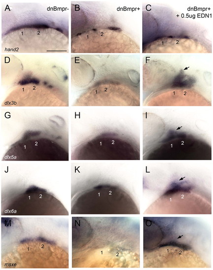
Edn1 rescues ventral patterning in BMP-deficient embryos. (A-O) Whole-mount in situ hybridization to detect expression of genes involved in DV arch patterning at 30 hpf, lateral views, anterior towards the left in controls (A,D,G,J,M), Tg(hsp70I:dnBmpr1a-GFP) transgenics heat shocked at 16-18 hpf (B,E,H,K,N) and heat-shocked transgenics injected with human recombinant EDN1 protein (C,F,I,L,O). hand2 (A-C) expression is unaffected, whereas dlx5a (G-I), dlx6a (J-L) and msxe (M-O) expression are restored by EDN1 protein in heat-shocked transgenics, and dlx3b (D-F) expression is induced throughout the DV extent of the arches. Arches 1 and 2 are numbered. Arrows indicate rescued gene expression. Scale bar: 100 μm. |
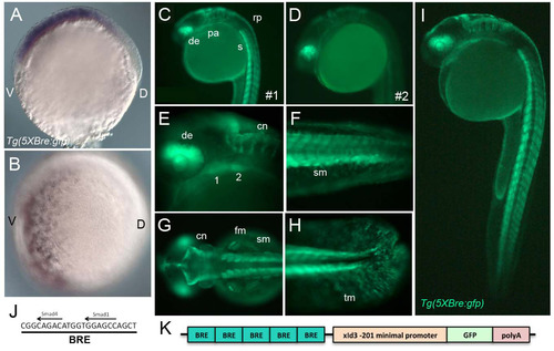
Tg(Bre:GFP) expression. (A,B) Whole-mount in situ hybridization for GFP mRNA at early gastrula stage (6 hpf), lateral (A) and ventral (B) views, dorsal towards the right. (C-I) Live images of Tg(Bre:GFP) expression, lateral views (except G, which is a dorsal view), anterior to the left. (C,D) Two stable Tg(Bre:GFP) transgenic lines (1 and 2), derived from independent insertion events, show similar expression patterns. (J) The BRE sequence includes putative Smad1- and Smad4-binding sites separated by five bases. (K) The Tg(Bre:GFP) transgenic construct includes five tandem Bres (blue-green), a minimal XId3 promoter (orange), GFP (green) and an SV40 polyA tail (red). 1, mandibular arch; 2, hyoid arch; cn, commissural neurons; de, dorsal eye; fm, fin muscle; pa, pharyngeal arches; sm, somite muscle; tm, tail mesenchyme. |
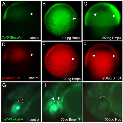
BMP signaling is sufficient and necessary for Tg(Bre:GFP) expression. (A-C) Whole-mount immunochemical staining for GFP protein at early gastrula stages, lateral views, ventral towards the left. (D-F) Whole-mount immunochemical staining for pSmad1/5/8 protein at early gastrula stages, lateral views, ventral towards the left. (A,D) Uninjected controls. (B,E) Embryos microinjected at the one-cell stage with 100 pg xBmp4 mRNA. (C,F) Embryos injected with 250 pg xBmp4. (G-I) Whole-mount immunochemical staining for GFP protein at 30 hpf, lateral views of the head, anterior towards the left. (G) Control embryo in which a DMSO-soaked bead (dashed line) was implanted behind the eye at 24 hpf. (H) Increased GFP surrounding a similarly implanted bead soaked in 10 µg/µl human recombinant BMP4/7. (I) Loss of GFP in an embryo implanted with a bead soaked in 500 μg/μl NOG. Abbreviations: cn, commissural neurons; D, dorsal; de, dorsal eye; m, margin; pa, pharyngeal arches; V, ventral. |
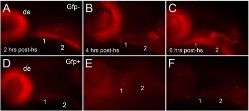
Loss of Smad1/5/8 phosphorylation in heat shocked Tg(hs:dnBmpr1a-GFP) embryos. (A-F) Immunochemical staining for pSmad1/5/8, lateral views, anterior towards the left. (A-C) GFP-negative, non-transgenic controls subjected to heatshock. (D-F) GFP-positive transgenics. (A,D) pSmad1/5/8 staining is already reduced in arches 1 and 2 in transgenics 2 hours post-heatshock (post-hs). (B,E) Staining is lost in the arches and in the eye by 4 hours post-hs. (C,F) 6 hours post-hs. 1, mandibular arch; 2, hyoid arch; de, dorsal eye. |
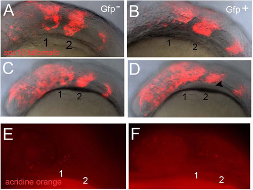
Loss of BMP signaling during arch formation does not disrupt NC migration or survival. (A-D) Live images of fluorescently labeled NC cells in Tg(sox10:tdTomato) transgenics, lateral views, anterior towards the left. Migratory streams of NC in the mandibular (1) and hyoid (2) arches appear identical in GFP- and GFP+ embryos. (E,F) Acridine Orange staining of apoptotic cells, lateral views, anterior towards the left, also reveals no difference in numbers of labeled cells in arches 1 and 2 between heat shocked hs-dnBmpr1a embryos and controls. |
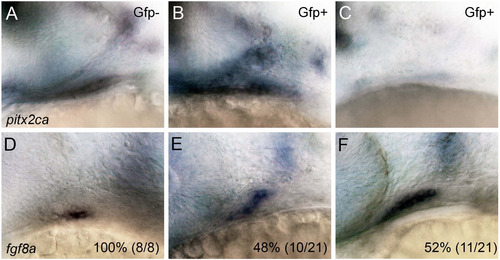
Loss of BMP signaling during arch formation disrupts expression of pitx2ca and fgf8a in the oral ectoderm. (A-F) Whole-mount in situ hybridization for pitx2ca (A-C) and fgf8a (D-F) at 30 hpf, lateral views, anterior towards the left. Heat shocked controls (A,D) and Tg(hsp70I:dnBmpr1a-GFP) embryos (B,C,E,F). Expression of pitx2ca is reduced and fgf8a expands in the stomodeum in the absence of BMP signaling. |
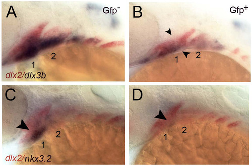
Loss of gene expression in the intermediate zone in heat shocked Tg(hs:dnBmpr1a-GFP) embryos. (A-D) Two-color in situ hybridization for dlx2a (red), dlx3b (blue in A,B) and nkx3.2 (blue in C,D) in heat-shocked controls (A,C) and Tg(hsp70I:dnBmpr1a-GFP) (B,D) embryos. |
Compound mutant/morphants reveal genetic interactions between dnBmpr1a and Edn1. (A-F) Alcian stained cartilages at 5 dpf, anterior towards the left. Compared with controls (A), ventral cartilage defects (particularly the size of Meckels cartilage, Mc) in heat-shocked heterozygous Tg(hsp70I:dnBmpr1a-GFP)+/ mutants (B,C) or embryos injected with 0.5 ng of Edn1 MO (D) are much less severe than in the double: Tg(hsp70I:dnBmpr1a-GFP)+/ plus 0.5 ng edn1-MO (E,F). |
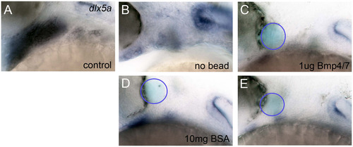
Beads soaked in human recombinant BMP4/7 do not rescue dlx5a expression in Edn1-deficient embryos. (A-E) Whole-mount in situ hybridization to detect expression of dlx5a at 30 hpf, lateral views, anterior towards the left in controls (A), edn1-MO injected embryos alone (B) or implanted with a BSA-soaked control bead (C) and two examples of edn1-MO injected embryos implanted with a BMP4/7 protein-soaked bead (C,E). Arches 1 and 2 are numbered. |