
- Title
-
Aberrant AKT activation drives well-differentiated liposarcoma
- Authors
- Gutierrez, A., Snyder, E.L., Marino-Enriquez, A., Zhang, Y.X., Sioletic, S., Kozakewich, E., Grebliunaite, R., Ou, W.B., Sicinska, E., Raut, C.P., Demetri, G.D., Perez-Atayde, A.R., Wagner, A.J., Fletcher, J.A., Fletcher, C.D., and Look, A.T.
- Source
- Full text @ Proc. Natl. Acad. Sci. USA
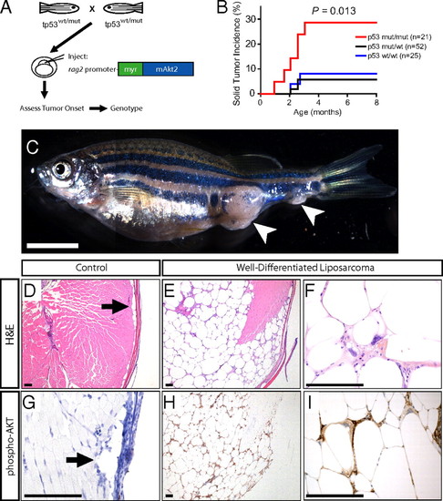
Constitutive Akt activation drives WDLPS in the zebrafish. (A) Experimental design. (B) Solid tumor incidence in p53 wild-type, heterozygous, or p53M214K homozygous mutant siblings injected with a rag2:myr-mAkt2 transgene at the one-cell stage. P value calculated via log-rank test. (C) Representative rag2:myr-mAkt2-injected zebrafish, which developed what appear to be two independent solid tumors. The animal shown was p53-homozygous mutant. (Scale bar, 5 mm.) Note that the image shown in (C) consists of merged adjacent photomicrographs. (D) Control H&E-stained zebrafish section. Arrow points to normal subcutaneous adipocytes. (E) Low-magnification view of an H&E section through the zebrafish shown in C, demonstrating a locally invasive mass consisting of well-differentiated adipocytes with significant variation in cell size. (F) High-magnification view of H&E section demonstrating a representative lipoblast scattered throughout these tumors, characterized by a multivacuolated cytoplasm and large hyperchromatic pleomorphic nuclei. (G) Phospho-AKT immunohistochemistry on a control zebrafish section. Arrow points to normal subcutaneous adipocytes, which lacked detectable pAKT staining. (H and I) Phospho-AKT immunohistochemistry on tumor sections from the zebrafish shown in C, revealing strong immunoreactivity for phospho-AKT in tumor cells of rag2:myr-mAkt2-transgenic zebrafish. (Scale bars, 100 μm.) |
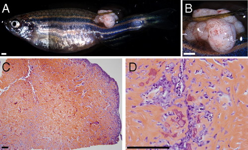
Osteosarcoma development in a rag2:myr-mAkt2-injected, p53-homozygous mutant zebrafish. (A and B) A p53-homozygous mutant zebrafish injected with the rag2:myr-mAkt2 expression construct developed a solid lobulated mass at the base of the dorsal fin. Note that the image shown in A consists of merged adjacent photomicrographs. (Scale bars, 1 mm.) (C and D) H&E-stained sections at low and high magnification, respectively, demonstrate a mass consisting predominantly of osteoid matrix interspersed with large malignant cells with pleomorphic nuclei, features that in humans are diagnostic of osteoblastic osteosarcoma. (Scale bars, 100 μm.) |

AKT pathway activation in primary human well-differentiated and dedifferentiated liposarcomas. (A–C) H&E staining of human lipoma, WDLPS, and DDLPS specimens. (D–F) Immunohistochemistry for Ser473-phosphorylated AKT in a representative lipoma and AKT-positive WDLPS and DDLPS clinical specimens. (Scale bar, 100 μm.) (G–I) Immunohistochemistry for phospho-S6 ribosomal protein (Ser235/236) in a representative lipoma, as well as AKT-positive WDLPS and DDLPS clinical specimens. (J and K) Quantitation of phospho-AKT and phospho-S6 immunohistochemistry in pure WDLPS, pure DDLPS, and in human tumors with mixed well-differentiated and dedifferentiated liposarcoma components. |