
- Title
-
The novel transmembrane protein Tmem2 is essential for coordination of myocardial and endocardial morphogenesis
- Authors
- Totong, R., Schell, T., Lescroart, F., Ryckebüsch, L., Lin, Y.F., Zygmunt, T., Herwig, L., Krudewig, A., Gershoony, D., Belting, H.G., Affolter, M., Torres-Vazquez, J., and Yelon, D.
- Source
- Full text @ Development
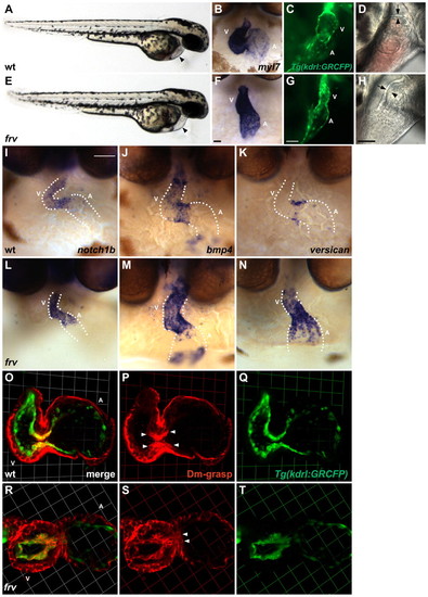
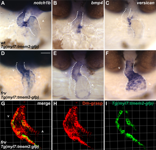
The frv mutation disrupts myocardial and endocardial morphogenesis. (A,E) Lateral views of wild-type (wt) and frv mutant zebrafish embryos at 48 hpf reveal mild pericardial edema in frv mutants (arrowheads). Other than their cardiac defects, frv mutants appear morphologically normal. (B,F) Frontal views depict myl7 expression in wild type (B) and in frv mutants (F) at 48 hpf. (C,G) Lateral views showing endocardial expression of Tg(kdrl:GRCFP) in wild type (C) and in frv mutants (G) at 52 hpf. (D,H) Lateral views at 52 hpf depict the close juxtaposition of the ventricular endocardium (arrowhead) and myocardium (arrow) in wild type (D) and their greater separation in frv mutants (H). (I-N) Frontal views depict expression of atrioventricular canal (AVC) markers in wild type (I-K) and in frv mutants (L-N) at 48 hpf. Dotted lines outline the chambers flanking the AVC. (O-T) Three-dimensional projections of selected confocal sections of wild-type (O-Q) and frv mutant (R-T) hearts expressing Tg(kdrl:GRCFP) (green) in the endocardium at 57 hpf. Immunofluorescence reveals Dm-grasp (red) throughout the myocardium and in the AVC endocardium (arrowheads). In frv mutants, Dm-grasp is also seen ectopically in the ventricular endocardium. Grids are 23 μm per segment. A, atrium; V, ventricle. Scale bars: 50 μm. |
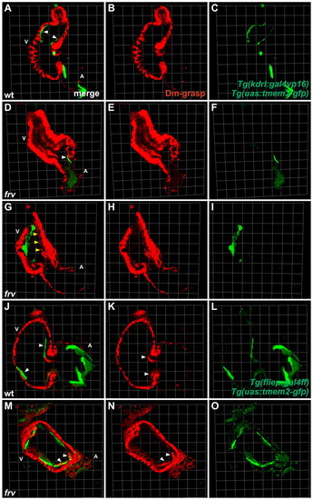
Endocardial expression of tmem2 rescues the frv mutant endocardium. Three-dimensional projections of selected confocal sections of wild-type (A-C,J-L) and frv mutant (D-I,M-O) zebrafish hearts expressing tmem2-gfp (green) in the endocardium at 57 hpf. Immunofluorescence indicates Dm-grasp localization (red). (A-I) Transient mosaic expression of tmem2-gfp is driven by injection of the transgenes Tg(kdrl:gal4vp16) and Tg(uas:tmem2-gfp). (J-O) Mosaic expression of tmem2-gfp is driven by stably integrated Tg(fliep:gal4ff) and Tg(uas:tmem2-gfp) transgenes. (A-C,J-L) Expression of tmem2-gfp in ventricular endocardium (A,J, arrowheads) does not affect wild-type development. (D-F) Ectopic Dm-grasp remains in the frv mutant ventricle when tmem2-gfp is expressed only in atrial endocardium (D, arrowhead). (G-I) Mosaic expression of tmem2-gfp in the ventricular endocardium of frv mutants suppresses ectopic Dm-grasp localization. Ventricular endocardium that does not express tmem2-gfp retains ectopic Dm-grasp (G, arrowheads). (M-O) Broad tmem2-gfp expression in the ventricular endocardium rescues ectopic Dm-grasp expression in an frv mutant. AVC endocardium still expresses Dm-grasp as in wild type (compare M,N with K, arrowheads). Endocardial expression of tmem2-gfp does not improve ventricular contractility, but does reduce the abnormal gap between the myocardium and endocardium. Grids are 23 μm per segment. |
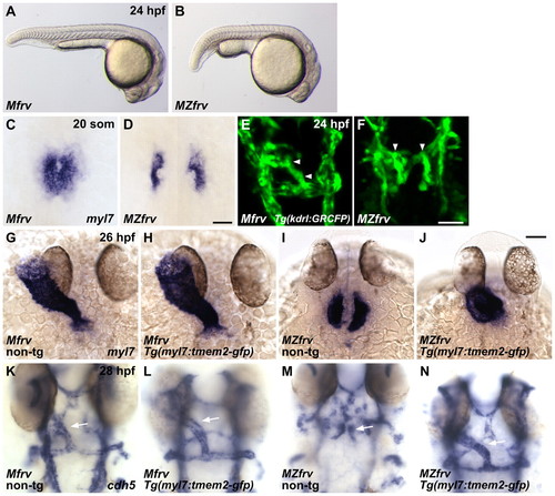
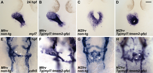
Maternal-zygotic frv mutants exhibit myocardial and endocardial fusion defects that are rescued by myocardial tmem2 expression. (A,B) Lateral views of zebrafish maternal (Mfrv) and maternal-zygotic (MZfrv) frv mutant siblings reveal body length and yolk extension defects in MZfrv embryos at 24 hpf. (C,D,G-J) Dorsal views depict myl7 expression at 20 somites (som) and 26 hpf. (E,F) Dorsal views depict endocardial expression of Tg(kdrl:GRCFP) (arrowheads) at 24 hpf. (K-N) Dorsal views depict endocardial expression of cdh5 (arrows) at 28 hpf In Mfrv embryos, expression of Tg(myl7:tmem2-gfp) has no effect on myocardial or endocardial morphogenesis (G,H,K,L), whereas in MZfrv embryos expression of Tg(myl7:tmem2-gfp) rescues both myocardial and endocardial fusion (I,J,M,N). Non-transgenic (non-tg) and transgenic siblings were distinguished by examination of fluorescence before fixation. Comparable results were obtained with two independent transgenic founders. Scale bars: 50 μm. EXPRESSION / LABELING:
PHENOTYPE:
|
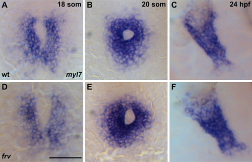
Cardiac fusion and heart tube elongation occur normally in frv mutants. Dorsal views, anterior to the top, depicting myl7 expression. (A,D) Cardiac fusion begins at the 18-somite stage (som) in both wild-type (wt) (A) and frv mutant (D) embryos. (B,E) By 20 som, cardiac fusion is complete in both wt (B) and frv mutant (E) embryos. (C,F) At 24 hpf, both wt (C) and frv mutant (F) embryos display an elongated heart tube. Similarly, examination of vmhc and amhc expression did not reveal defects in frv mutants at these stages (data not shown). Scale bar: 50 μm. EXPRESSION / LABELING:
PHENOTYPE:
|
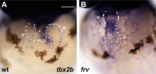
Expanded expression of tbx2b in frv mutants. (A,B) Frontal views depicting expression of tbx2b at 48 hpf in wt (A) and frv mutant (B) embryos. Dotted lines outline the chambers flanking the AVC; brackets demarcate the extent of tbx2b expression. Expression of tbx2b is normally restricted to AVC myocardium (A), but it expands into the cardiac chambers in frv mutants (B). A, atrium; V, ventricle. Scale bar: 50 μm. |
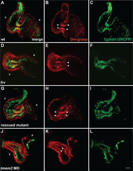
mRNA rescue and morpholino phenocopy of the frv mutant phenotype. Three-dimensional projections of selected confocal sections, as in Fig. 1O-T, of hearts expressing Tg(kdrl:GRCFP) (green) in the endocardium at 57 hpf. (A-C) In wt embryos, immunofluorescence recognizes Dm-grasp (red) throughout the myocardium and in the AVC endocardium (arrowheads, B). (D-F) In frv mutants, Dm-grasp is also seen ectopically in the ventricular endocardium (arrowheads, E). (G-I) Injection of frv mutants with tmem2 mRNA can eliminate ectopic Dm-grasp from the ventricular endocardium (see Table S1 in the supplementary material). In this example of a rescued mutant, endocardial Dm-grasp is restricted to the AVC (arrowheads, H). (J-L) Injection of wt embryos with an anti-tmem2 MO can result in ectopic Dm-grasp in the ventricular endocardium (arrowheads, K), as well as dysmorphic chambers and reduced ventricular contractility. Out of 117 MO-injected embryos, 48 exhibited frv-like characteristics. Although MO injection did not fully recapitulate the extent of ectopic Dm-grasp observed in frv mutants, it is notable that the MO could cause Dm-grasp localization where it is not observed in wt embryos. We presume that the degree of MO phenocopy reflects incomplete knockdown of tmem2. Scale bar: 20 μm. |
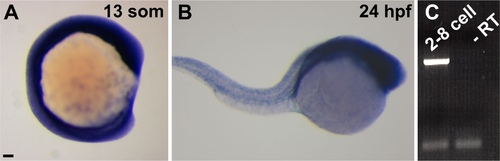
tmem2 is broadly and maternally expressed. (A,B) Lateral views of wt embryos, dorsal up, anterior to the right. (A) In situ hybridization with a probe corresponding to bases 2446-3524 of the tmem2 coding sequence indicates ubiquitous expression at 13 som. (B) At 24 hpf, tmem2 is still broadly expressed throughout the embryo, with highest expression levels in the anterior. (C) RT-PCR using tmem2-specific primers and cDNA from wt embryos at the 2- to 8-cell stage indicates that tmem2 is maternally provided. The 1100 bp product in the first lane is a fragment of tmem2 cDNA. The -RT control lane shows no amplification when reverse transcriptase is excluded from the reverse transcription reaction. The lower bands in both lanes are primer dimers. Scale bar: 50 μm. |
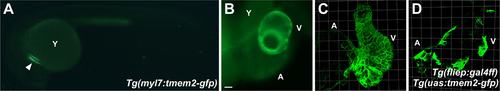
Expression patterns of the Tg(myl7:tmem2-gfp), Tg(fliep:gal4ff) and Tg(uas:tmem2-gfp) transgenes. (A) Lateral view of a live embryo at 24 hpf expressing the stably integrated transgene Tg(myl7:tmem2-gfp), in which tmem2-gfp expression is driven by the myl7 promoter (Huang et al., 2003). Expression is detected only in the myocardium (arrowhead). Faint autofluorescence is visible in the yolk (Y). (B) Ventricular view of a live heart at 57 hpf expressing the stably integrated transgene Tg(myl7:tmem2-gfp) throughout the myocardium. (C) Three-dimensional confocal reconstruction of a live heart at 57 hpf expressing the stably integrated transgene Tg(myl7:tmem2-gfp) demonstrates localization of GFP to the plasma membrane. (D) Three-dimensional confocal reconstruction of a live heart at 57 hpf expressing the stably integrated transgenes Tg(fliep:gal4ff) and Tg(uas:tmem2-gfp) demonstrates mosaic expression in the ventricular and atrial endocardium. This degree of mosaicism was adequate for examining rescue of the ventricular endocardium in zygotic frv mutants (see Fig. 3J-O). However, evaluation of whether endocardial expression of tmem2 can rescue cardiac fusion in MZfrv mutants will require future development of less mosaic Tg(uas:tmem2-gfp) lines. Scale grids are 23 μm per segment. Scale bar: 50 μm. |
Myocardial expression of tmem2 does not rescue frv mutants. (A-F) Frontal views depicting expression of AVC markers at 48 hpf (as in Fig. 1). All embryos shown carry the Tg(myl7:tmem2-gfp) transgene, as confirmed by examination of fluorescence prior to fixation. (A-C) Expression of Tg(myl7:tmem2-gfp) does not alter the phenotype of wt embryos (compare with Fig. 1I-K). (D-F) Expression of Tg(myl7:tmem2-gfp) does not alter the phenotype of frv mutant embryos (compare with Fig. 1L-N). (G-I) Three-dimensional projections of selected confocal sections of an frv mutant heart expressing Tg(myl7:tmem2-gfp) (green) in the myocardium at 57 hpf. Immunofluorescence reveals no effect of the transgene on ectopic localization of Dm-grasp (red) (compare with Fig. 1R-T). Comparable results were obtained with two independent transgenic lines. Scale grids are 23 μm per segment. Scale bar: 50 μm. |
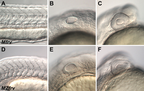
MZfrv mutants exhibit multiple morphological defects. (A-F) Lateral views of Mfrv and MZfrv mutant siblings at 24 hpf reveal pleiotropy in MZfrv mutant embryos, including slightly curved somites (D), a lack of otoliths (E), and a small head (F), potentially owing to failure of brain ventricle inflation. Scale bar: 50 μm. PHENOTYPE:
|
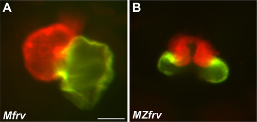
MZfrv mutant hearts are bifurcated. Ventral views of hearts at 48 hpf. (A) Immunofluorescence employing the MF20 and S46 antibodies demonstrates that Mfrv mutant embryos develop normally looped hearts with a distinct ventricle (red) and atrium (yellow). (B) In MZfrv mutant embryos, the heart is bifurcated with subdivided ventricular (red) and atrial (yellow) chambers. Scale bar: 50 μm. |
Myocardial expression of tmem2 rescues myocardial and endocardial fusion defects in MZfrv mutants. (A-D) Dorsal views depict myl7 expression at 24 hpf. (E-H) Dorsal views depict endocardial expression of cdh5 (arrowheads) at 24 hpf. (A,B,E,F) Expression of Tg(myl7:tmem2-gfp) has no effects on myocardial or endocardial morphogenesis in Mfrv embryos. (C,D) Expression of Tg(myl7:tmem2-gfp) rescues myocardial fusion in MZfrv embryos. The rate of cardiomyocyte movement varies among MZfrv mutants, such that some mutants exhibit contact between contralateral populations of cardiomyocytes at 24 hpf (C), whereas others do not exhibit any contact even at 26 hpf (see Fig. 4I). Even so, expression of Tg(myl7:tmem2-gfp) always results in significant acceleration of myocardial morphogenesis in MZfrv mutants. In this example (D), myocardial fusion is complete and myocardial tube extension has initiated. (G,H) Expression of Tg(myl7:tmem2-gfp) rescues endocardial fusion in MZfrv embryos. In this example (H), endocardial fusion is complete and endocardial tube extension has begun. Non-transgenic (non-tg) and transgenic siblings were distinguished by examination of fluorescence before fixation. Comparable results were obtained with two independent transgenic founders. Scale bar: 50 μm. |

Unillustrated author statements PHENOTYPE:
|