
- Title
-
Mediator subunit 12 is required for neutrophil development in zebrafish
- Authors
- Keightley, M.C., Layton, J.E., Hayman, J.W., Heath, J.K., and Lieschke, G.J.
- Source
- Full text @ PLoS One
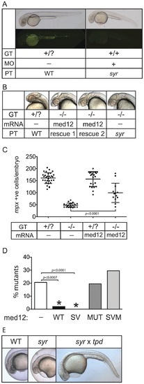
Genetic validation of V1046D mutant Med12 underpinning syr phenotype. (A) Phenocopy of syr by injection of Med12 morpholino; phenotype (PT), genotype (GT) and morpholino (MO) injection are indicated. The upper panel are bright field photos and the lower panels show WT and syr on a Tg(mpx:EGFP) background for enumeration of mpx expressing cells. (B) Rescue of syr neural phenotype with wild-type Med12; phenotype (PT), genotype (GT) and mRNA injected. (C) Rescue of myeloid defect in syr by overexpression of wild-type Med12 mRNA; genotype (GT) and mRNA injected are indicated. (D) Rescue of syr with wild-type (WT) Med12 and Med12 containing the two sequence variations, N1763S and a 12 bp deletion but not the V1046D mutation (SV). Rescue does not occur with injection of V1046D Med12 (MUT) or Med12 containing the two sequence variations in addition to the V1046D mutation (SVM). (E) Non-complementation of syr with trapped (tpd). Rescued mutants were PCR genotype confirmed. |

Hematopoiesis defects in syr. Myelomonocytic markers were examined at 23–28 hpf by WISH (A–N); T lymphocyte and thymic epithelium markers were examined at 3.5 dpf in syr and wt (O–R); staining of the thrombocyte marker, cd41 at 3 dpf (S–T). WISH embryos are representative of e4 (median = 21) examples. EXPRESSION / LABELING:
PHENOTYPE:
|
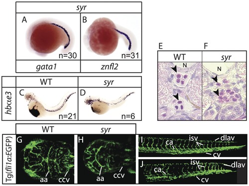
Erythropoiesis proceeds normally in syr. Examination of early erythroid markers at 17–19 hpf in syr (A–B); staining of embryonic globin in wt and syr at 48 hpf (C–D); transverse sections of WT and syr embryos at 3 dpf, counterstained with hematoxylin and eosin; notochord (N) and neutrophils (arrowheads) are indicated (E–F); syr crossed with Tg(fli1a:EGFP) shows close to normal vasculature in both head (G–H) and tail (I–J). Heads are dorsal view with anterior to left, WT on the left (G) and syr on the right (H); aa (aortic arches) and ccv (common cardinal vein) are indicated. Tails are lateral view, anterior to left and ca (caudal artery), cv (caudal vein), dlav (dorsal longitudinal anastomotic vessel) and isv (intersomitic vessels) are indicated. EXPRESSION / LABELING:
PHENOTYPE:
|
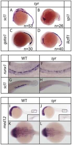
Stages of hematopoiesis in syr. Early hematopoietic markers are expressed normally in syr at 17–19 hpf (A–D) (note that Figs. 4A and 5C are the same; Fig. 4A/5C is included here for completeness). Definitive hematopoiesis is initiated in syr indicated by runx1 expression at 28 hpf (E–F). Caudal hematopoietic tissue fails to develop in syr indicated by lack of scl1 expression at 3 dpf (G–H). Med12 is expressed in WT and syr with insets showing staining in the hematopoietic ICM region (I–J); dorsal view of staining (K–L). Unless otherwise stated, WISH embryos are representative of e10, and d46, examples. EXPRESSION / LABELING:
PHENOTYPE:
|
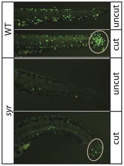
Residual neutrophils can migrate in syr. Top panels: WT prior to tail snip (uncut) and 8 h post transection (cut); Bottom panels: Syr prior to tail snip (uncut) and 8 h post transection (cut). Neutrophils at the wound margin have been circled. |