
- Title
-
miR-196 regulates axial patterning and pectoral appendage initiation
- Authors
- He, X., Yan, Y.L., Eberhart, J.K., Herpin, A., Wagner, T.U., Schartl, M., and Postlethwait, J.H.
- Source
- Full text @ Dev. Biol.
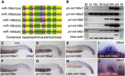
Sequence and expression of mir196 genes. (A) Alignment of mature mir196 encoded by five mir196 genes. (B) Expression of mir196 paralogs studied by RT-PCR. Beta-actin (bactin1) was used as control for contaminating genomic DNA (ctrl lane). M, size marker; 1c, 1 cell stage; 16c, 16 cell stage; 5 h, 5 hpf (hours post-fertilization); bud, bud stage, about 10 hpf; 24 h, 24 hpf; ctrl, genomic DNA control. (C–H) Whole mount in situ hybridization for mir196 primary transcripts showed expression in the tail bud and neural tube. (H) Linked nucleic acid (LNA) probe for miR-196a showed an expression pattern similar to the primary transcript. (I, J) LNA probes for mir196a in the pectoral fin bud at 30 and 48 hpf. cns, central nerve system; pf, pectoral fin. |
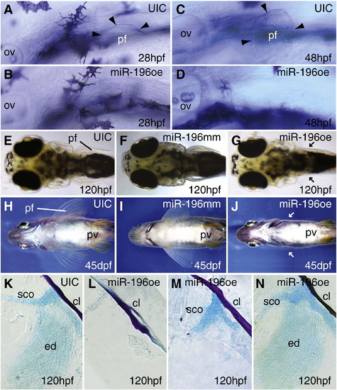
miR-196 overexpression inhibits pectoral fin initiation. (A, C) Pectoral fin buds are readily detectable at 28 hpf and 48 hpf in normally developing uninjected control animals, but embryos overexpressing miR-196 (B, D) showed no evidence of a pectoral fin bud. Arrowheads mark the edges of pectoral fin buds. (E, F, H, I) Uninjected controls or miR-196 mismatch injected controls showed normal pectoral fins by 5 and 45 days post-fertilization (dpf), but larvae overexpressing miR-196 had not recovered pectoral fins by 5 dpf and became paraplegic adults (G, J, arrows point to missing pectoral fins). Pelvic fins were normal in fish with pectoral fin defects (J). (K–N) Dissected pectoral fins of 5 dpf larvae stained with Alcian blue for cartilage and Alizarin red for bone showed skeletal defects ranging from absence of the endochondral disc and scapulocoracoid (L, 42.3%, 234 total fins) to missing the endochondral disc but having part of scapulocoracoid (M, just 6%) to normal (N, 51.7%,). Abbreviations: cl, cleithrum; ed, endochondral disc; ov, otic vesicle; pf, pectoral fin or bud; pv, pelvic fin; sco, scapulocoracoid. |
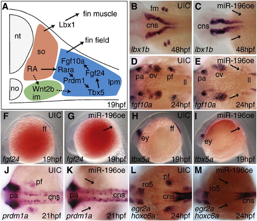
miR-196 duplex injection inhibits expression of zebrafish fin development genes. (A) Model of the pectoral fin developmental pathway (after Mercader (2007)). In situ hybridization for lbx1b (B, C), ifgf10a (D, E), fgf24 (F, G), tbx5a (H, I), prdm1a (J, K), hoxc6a (L, M), on controls (B, D, F, H, J, L) and miR-196 overexpression (oe) animals (C, E, G, I, K, M) at ages indicated in the lower right of each panel. Arrowheads show the presumptive pectoral fin region in miR-196 duplex-injected animals. Results showed that miR-196 acts at or before the earliest stages of fin bud initiation. Abbreviations: cns, central neural system; ff, fin field; fm, pectoral fin muscle; im, intermediate mesoderm; ll, lateral line primordium; no, notochord; nt, neural tube; ov, otic vesicle; pa, pharyngeal arch; pf, pectoral fin bud; ro5, rhombomere 5. |
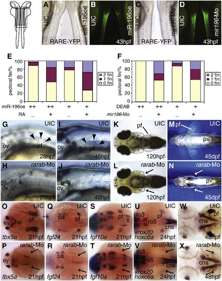
miR-196 activity modulates RA signaling. (A) Bright field microscopy of a 43 hpf uninjected control transgenic RA signaling reporter individual and a miR-196 injected 43 hpf RA-reporter embryo oriented back-to-back as in the insert at the left of part (A), with the boxed region blown up in A–D. (B) The animals in (A) was viewed in fluorescence microscopy. The intensity of green fluorescence is proportional to the level of RA signaling, and was greatly reduced after miR-196 overexpression. (C) Bright field and (D) fluorescence views of a control (left) and a miR-196-morpholino injected 43 hpf embryo (right). The elevated expression of YFP found after miR-196 knockdown is opposite to the phenotype found after miR-196 overexpression. (E, F) Rescue experiments. (E) Although injecting cleavage embryos with 5 nL of miR-196 duplex and treating them with DMSO as controls (++) caused a greater fraction of animals to develop without pectoral fins (89%, n = 63) than injections with 1 nL (+) (78%, n = 76), both can be rescued by RA treatment (+) (48%, n = 81 and 28% n = 61 without fin respectively). This experiment was repeated four times. (F) Treating embryos with 1 × 10⁻⁵ M (++) or 5 × 10⁻⁶ M (+) DEAB to decrease RA levels resulted in all (100%, n = 31) or most of the animals (87.5%, n = 16) lacking pectoral fins respectively, but first injecting cleavage embryos with 1 nL of 3 mM mir196-Mo (+) to diminish the inhibition of RA signaling before treating embryos with DEAB, led to partial rescue of the fin phenotype (just 53%, n = 51 or 54%, n = 48, respectively). Compared to controls (G, I, K, M), rarab-Mo injection (H, J, L, N) inhibited pectoral fin outgrowth already by 28 hpf (G, H) and this phenotype was maintained through 48 hpf (I, J), 120 hpf (K, L), and into adulthood (M, N) verifying the specificity of the effect. Arrowheads mark the edge of the pectoral fin and arrows direct attention to missing pectoral fins and buds. (O–V) rarab-Mo inhibited expression of the pectoral fin genes tbx5a (O, P), fgf24 (Q, R), fgf10a (S, T), and hoxc6a (U, V), as well as the fin muscle marker gene lbx1b (W, X) specifically in the fin field region while leaving other expression domains intact, the same result as from miR-196 injection. Arrows: missing pectoral fin buds. cns, central neural system; ey, eye; he, heart; im, intermediate mesoderm; ll, lateral line primordium; ov, otic vesicle; pa, pharyngeal arch; pf, pectoral fin; pv, pelvic fin; ro3, -5, rhombomere3, -5. EXPRESSION / LABELING:
|
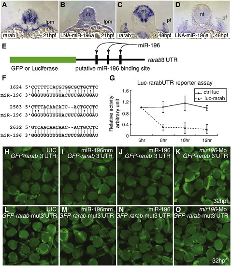
rarab is a target of miR-196. (A–D) In situ hybridization on tissue sections for rarab (A, C) and miR-196 (using LNA probe, B, D) at 21 hpf (A, B) and 48 hpf (C, D) shows that rarab and miR-196 are co-expressed in the lateral plate mesoderm at 21 hpf (A, B) and medially in the pectoral fin bud at 48 hpf (C, D). (E–O) Reporter assays. (E) Reporter assay constructs used luciferase in (G) and GFP in (H–K). (F) The 3′UTR of rarab has three predicted binding sites for miR-196 (nucleotide position according to NM_131399, binding sites were retrieved or predicted from miRBase.com and MicroInspector). (G) miR-196 can interfere with expression of a luciferase reporter construct bearing the 3′UTR of rarab compared to control. This experiment was repeated three times. (H–K) GFP-rarab 3′UTR mRNA was injected into early cleavage stage embryos either by itself (H) or co-injected with miR-196 mismatch (I), with miR-196 duplex (J), or with mir196-Mo (K). miR-196 depressed fluorescence but mir196 knockdown enhanced fluorescence, as predicted by the hypothesis that miR-196 acts directly on the 3′UTR of rarab. This experiment was repeated four times. (L–O) The three predicted miR-196 binding sites were removed from the GFP-rarab 3′UTR construct to make a GFP-rarab-mut3′UTR. GFP-rarab mut3′UTR mRNA was injected into early cleavage stage embryos either by itself (L) or co-injected with miR-196 mismatch (M), with miR-196 duplex (N), or with mir196-Mo (O). miR-196 did not depress fluorescence (N) and mir196 knockdown did not enhance fluorescence (O) from this mutant construct. This experiment was repeated three times. lpm; lateral plate mesoderm; pf, pectoral fin bud. EXPRESSION / LABELING:
|
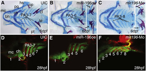
miR-196 regulates pharyngeal arch segmentation via pharyngeal endoderm. (A–C) Zebrafish embryos injected with miR-196 duplex or mir196-Mo were raised to 6 dpf and stained with Alcian blue for cartilage and Alizarin red for bone. (A) Control larvae had seven pharyngeal arches, including Meckels and ceratohyal cartilages (in PA1 and PA2) and five branchial arches (in PA3–7). (B) Animals overexpressing miR-196 lacked one branchial arch. Some animals with four branchial arches (insert) possessed a skeletal element attached to the last arch (arrows), suggesting that the missing arch was the one anterior to the tooth-bearing pharyngeal arch-7. (C) Injection of mir196-Mo resulted in animals with extra branchial arches. Some animals with seven pharyngeal arches (insert) displayed extra skeletal elements attached to pharyngeal arch-7 (arrows), suggesting that the extra arch was the one just anterior to PA7. (D–F) Transgenic animals expressing GFP in the neural crest (green) that were either overexpressing (E) or knocked down (F) for miR-196 showed defective segmentation of endodermal pouches stained with zn-8 antibody (red) and pharyngeal arches (green) consistent with observed alterations in arch numbers. Abbreviations: bh, basihyal; ch, ceratohyal; cl, cleithrum; hs, hyosymplectic; mc, Meckels cartilage; op, opercle; pa, pharyngeal arch; pf, pectoral fin; pq, palatoquadrate; te, teeth. |

Over- and under-expression of miR-196 disrupts patterning of the axial skeleton. (A) Normal axial skeleton stained for cartilage (Alcian blue) and bone (Alizarin red) showing 4, 10, 15 and 3 vertebrae in the Weberian apparatus (region W), rib-bearing precaudal vertebra (region P), caudal vertebra (region C) and caudal fin vertebra (region T), respectively. (B, D) Overexpression of mir196 showing deletion of one Weberian vertebra and three ribs with their precaudal vertebrae. (C, D) Morpholino knockdown of miR-196 resulted in extra ribs and extra rib-bearing precaudal vertebrae. (D) Skeletal element counts after miR-196 manipulation showing reciprocal effects of overexpression and knockdown in the rostral axial skeleton. Asterisks indicate statistical significance compared to UIC (uninjected control) (Student T-test, p < 0.001). UIC, n = 79; miR-196oe (overexpression), n = 56; mir196-Mo (miR-196 knockdown), n = 122. (E, F, I, J) Weberian apparatus sketch (after (Grande and Young, 2005)) and skeleton staining, lateral and dorsal views, respectively. (G, H, K, L) Overexpression of miR-196, lateral (G, H) and ventral (K, L) views of the Weberian apparatus from four different individuals. In wild-type fish, the Weberian apparatus has four highly modified vertebrae, each with bone and cartilage morphologies that specify their identity (E, F, I, J). Typically, the first vertebra (v1) has a pair of anterior-laterally projecting bones called the lateral process-1 (lp1) and dorsally, a small bone called intercalarium (in) that contacts with vertebra-2 (v2). V2 normally has a pair of laterally projecting bones (the lateral process-2, lp2) on each side. Lp2 is larger than lp1 (E, I). Vertebra-3 (v3) has a pair of lateral tripus (tr) bones, a triangular shaped bone, and neural arch-3 (na3) dorsally. Vertebra-4 (v4) has a pair of highly modified ribs (r4) protruding ventral-laterally that bears os suspensorium bones ventrally and dorsally, a neural arch-4, on top of which, supranueral-4 (sn4) usually fuses anteriorly with supranueral-3 (sn3) on v3. Precaudal vertebrae are defined by their parapophyses, which support ribs, and their lack of hemal arches and hemal spines, which are found on v15-v28 (Bird and Mabee, 2003). Treated animals are discussed in the text. Abbreviations: #1, #2, #3, vertebrae numbers from anterior without regard to identity; in, intercalarium; lp1, lp2, lateral processes; na3, na4, neural arches; os, os suspensorium; r4, r5, ribs; tr, tripus; v1, v2, v3, v4, v5, v7 vertebrae with identities. PHENOTYPE:
|

ZFIN is incorporating published figure images and captions as part of an ongoing project. Figures from some publications have not yet been curated, or are not available for display because of copyright restrictions. |
|
ZFIN is incorporating published figure images and captions as part of an ongoing project. Figures from some publications have not yet been curated, or are not available for display because of copyright restrictions. |
|
ZFIN is incorporating published figure images and captions as part of an ongoing project. Figures from some publications have not yet been curated, or are not available for display because of copyright restrictions. |
|
ZFIN is incorporating published figure images and captions as part of an ongoing project. Figures from some publications have not yet been curated, or are not available for display because of copyright restrictions. |
|
ZFIN is incorporating published figure images and captions as part of an ongoing project. Figures from some publications have not yet been curated, or are not available for display because of copyright restrictions. EXPRESSION / LABELING:
PHENOTYPE:
|
|
ZFIN is incorporating published figure images and captions as part of an ongoing project. Figures from some publications have not yet been curated, or are not available for display because of copyright restrictions. PHENOTYPE:
|
|
ZFIN is incorporating published figure images and captions as part of an ongoing project. Figures from some publications have not yet been curated, or are not available for display because of copyright restrictions. |
|
ZFIN is incorporating published figure images and captions as part of an ongoing project. Figures from some publications have not yet been curated, or are not available for display because of copyright restrictions. EXPRESSION / LABELING:
|
|
ZFIN is incorporating published figure images and captions as part of an ongoing project. Figures from some publications have not yet been curated, or are not available for display because of copyright restrictions. PHENOTYPE:
|
Reprinted from Developmental Biology, 357(2), He, X., Yan, Y.L., Eberhart, J.K., Herpin, A., Wagner, T.U., Schartl, M., and Postlethwait, J.H., miR-196 regulates axial patterning and pectoral appendage initiation, 463-77, Copyright (2011) with permission from Elsevier. Full text @ Dev. Biol.