
- Title
-
Zebrafish cardiac development requires a conserved secondary heart field
- Authors
- Hami, D., Grimes, A.C., Tsai, H.J., and Kirby, M.L.
- Source
- Full text @ Development
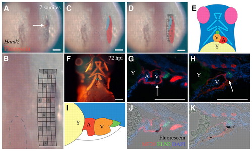
Arterial pole progenitors at the 7-somite stage. (A) Dorsal view of a 7-somite zebrafish embryo, cranial to the top. Photoactivated fluorescein, labeled immunohistochemically (red, arrow), can be identified relative to the hand2 expression field (blue/black). (B) A magnification of the image in A with a grid superimposed on the region targeted for uncaging to show experimental division of the heart field in the right anterior lateral plate mesoderm (ALPM). Gray circles represent ~4-cell clusters that were labeled in the mesodermal heart field. In this specimen, cells were labeled in grid zone F (red). The dashed line highlights the cranial tip of the notochord. (C) Dorsal view of a 7-somite embryo with a map showing the cardiogenic field (red) and arterial pole progenitors within that field (blue), based on Table 1. (D) Overlay of the grid from B and map from C. (E) Diagram of the ventral view of a zebrafish embryo at 72 hpf, with cranial indicated by the eyes (pink) and caudal indicated by the yolk (yellow). The branchial muscles (light blue) are cranial to the bulbus arteriosus (green), ventricle (orange) and atrium (red). (F) Zebrafish in similar orientation as in E. Progeny of cells labeled in grid zone F (black) at the 7-somite stage are shown relative to the bulbus arteriosus (anti-Eln2, green), and striated muscle (MF20, red). (G,H,J,K) Sagittal sections of 72 hpf zebrafish, with cranial to the left, showing progeny of the photoactivated population at grid zone F in the arterial pole (arrows). (I) Diagram showing the various parts of the heart seen in sagittal sections. Caudal is indicated by the yolk (yellow) and cranial by the bulbus arteriosus (green). A, atrium; V, ventricle; Y, Yolk. Scale bars: 10 μm. |
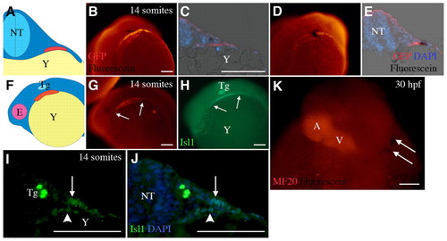
Cells that contribute to the arterial pole remain outside the early heart tube. (A) Diagram of a transverse section of a 14-somite embryo, with dorsal to the top, showing the neural tube (NT, light blue) with differentiating myocardium (red) in the ALPM overlying the yolk (Y, yellow). (B) Whole-mount 14-somite embryo in caudal view, dorsal to top. Labeled cells from grid zone F (black) were observed in the splanchnic mesoderm of Tg(cmlc2::GFP) zebrafish that expresses GFP in differentiated cardiomyocytes (orange). (C) Transverse section oriented as in A showing labeled cells (black) relative to differentiating myocardium (red). (D,E) Cells from grid zone B (black) can be seen in the differentiated myocardium (red) at 14 somites in whole mount (D) and in section (E). (F) Diagram of a 14-somite embryo from the left side, dorsal to top, showing the position of the cardiac progenitors (red), the eye (E, pink), the trigeminal placode (Tg, light blue) and the yolk (yellow). (G,H) A 14-somite embryo oriented as in F, showing Isl1 (H, green) and cardiomyocytes (G, orange). The trigeminal placode and the non-differentiating portion of the heart field are Isl1 positive. Arrows indicate the anterior and posterior extent of cmlc2::GFP or Isl1 signal in G and H, respectively. (I,J) Transverse sections of embryos at the 14-somite stage at the level of the trigeminal placode. Isl1 (green) can be barely detected in the nuclei (blue) of differentiating myocardium (arrowhead) but more brightly in the nuclei of cells in the adjacent splanchnic mesoderm (arrow). (K) Dorsal view of a 30 hpf zebrafish. Cells uncaged from grid zone F at the 7-somite stage (black, arrows) can be seen at some distance from the heart tube (orange). A, atrium; V, ventricle. Scale bars: 10 μm. |
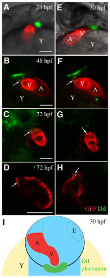
Cells in the pericardial wall are incorporated into the developing heart tube. (A,E) Cells were labeled with DiI (pseudocolored green) adjacent to the arterial pole of the heart tube (pseudocolored red) in Tg(cmlc2::GFP) zebrafish at (A) 24 hpf and (E) 30 hpf. Pseudocoloring improved the visibility of DiI-labeled cells. Zebrafish are shown from the left side, cranial to the top. (B,C,F,G) Labeled cells were observed in the GFP-positive myocardium at (B,F) 48 hpf and (C,G) 72 hpf. Zebrafish are shown from the right side, cranial to the top. (D,H) Optical sections of 72 hpf zebrafish hearts from the right side, cranial to the top, confirming that DiI-labeled cells were incorporated into the myocardium. (I) Diagram showing the approximate area of labeled cells (green) in the pericardial wall (black line) when viewed dorsally at 30 hpf. The heart tube is shown (red). Dashed lines indicate eyes (E). A, atrium; V, ventricle; Y, yolk. Scale bars: 10 μm. |
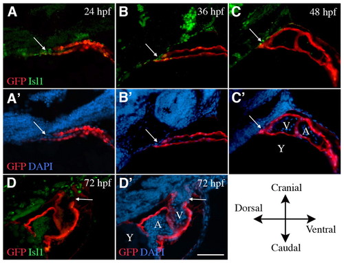
Isl1 is expressed by cells that are being incorporated into the arterial pole up to 48 hpf. Sagittal sections of Tg(cmlc2::GFP) zebrafish, cranial to the top, dorsal to the left. (A-C′) Isl1 (green) is expressed in cells adjacent to and within the arterial pole (arrows) at 24, 36 and 48 hpf during addition of the second heart field myocardial cells to the heart tube (GFP, red). (D,D′) By 72 hpf, Isl1 is absent from the myocardium and is expressed in the cranial-most portion of the smooth muscle of the bulbus arteriosus. A, atrium; V, ventricle; Y, yolk. Scale bar: 5 μm. |
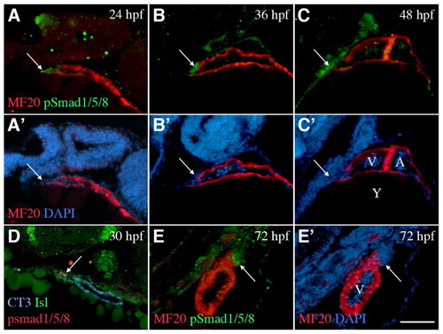
Phosphorylated Smad1/5/8 is present in cells being incorporated into the arterial pole up to 72 hpf. Sagittal sections of wild-type zebrafish, cranial to the top, dorsal to the left. (A-C′) pSmad1/5/8 is detected in cells adjacent to and within the arterial pole (arrows) at 24, 36 and 48 hpf during addition of the second heart field myocardial cells to the heart tube (MF20). (D) pSmad1/5/8 (red) is expressed in a subset of Isl1-positive cells (green) closest to the myocardium (CT3) at 30 hpf. (E,E′) By 72 hpf, pSmad1/5/8 (green) is expressed in the myocardium and the smooth muscle (red) of the bulbus arteriosus. A, atrium; V, ventricle; Y, yolk. Scale bar: 5 μm. |
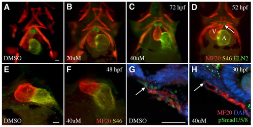
The absence of Bmp signaling induces an expansion of the bulbus arteriosus at the expense of the ventricle. (A-C) Ventral view of 72 hpf zebrafish treated with dorsomorphin (B,C) or DMSO carrier (A) from 24 to 72 hpf. (D) The bulbus arteriosus can be detected at 52 hpf as shown by anti-Eln2 labeling. A, atrium (S46); V, ventricle (MF20). (E,F) Ventral view of 48 hpf embryos. Dorsomorphin treatment (F) from 24 to 48 hpf leads to smaller ventricles (red) at 48 hpf. (G,H) Sagittal sections of embryos with dorsal to the top. Dorsomorphin treatment from 24 to 30 hpf (H) results in a significant loss of phosphorylation of Smad1/5/8 (green nuclei) at the arterial pole (arrow). Scale bars: 5 μm. |
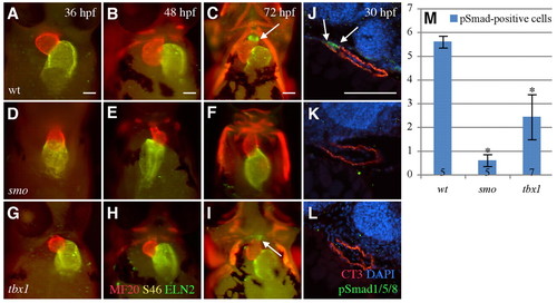
Zebrafish tbx1 and smo mutants display arterial pole defects. (A-C) Ventral view of whole-mount wild-type zebrafish, with cranial to the top. The heart is looped by 36 hpf and the bulbus arteriosus (Eln2) is formed by 72 hpf. Ventricle (MF20, red); atrium (S46, yellow). (D-I) In smo (D-F) and tbx1 (G-I) mutants, cardiac development is impaired. (J) Sagittal sections from the left show pSmad1/5/8 (green and arrows) near the arterial pole of the wild-type heart (CT3) at 30 hpf. (K,L) In the smo (K) and tbx1 (L) mutants, pSmad1/5/8 is significantly reduced. (M) Quantification of pSmad1/5/8-positive cells in wild-type versus smo and tbx1 mutants. Numbers in bars indicate the number of fish sampled. *, P=1.23×10⁻³. Error bars indicate s.d. Scale bars: 5 μm. |
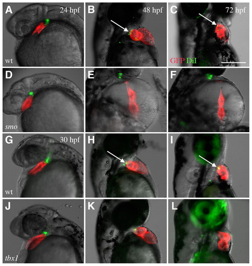
Labeled cells in the pericardial wall are not incorporated into the developing heart in smo and tbx1 mutants. (A-C,G-I) Cells labeled with DiI (pseudocolored green) in the branchial region are incorporated into the myocardium (pseudocolored red) in wild-type zebrafish hearts after labeling at 24 hpf (A-C) and 30 hpf (G-I). DiI colocalized with the myocardium in wild-type fish at 48 and 72 hpf (arrows). (D-F,J-L) This incorporation of cells does not occur in most smo (D-F) or tbx1 (J-L) mutants. The green cells that appear to overlap with red cardiac cells (K) do not overlap with the myocardium 24 hours later (L). All embryos expressed cmlc2-GFP. Scale bar: 25 μm. |
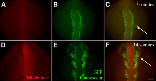
Arterial pole progenitors are not derived from neural crest. Ventral view, with cranial to top, of zebrafish after photoactivation of cells at the 7 somite stage. (A-C) When cells in grid zone F were labeled using caged fluorescein:rhodamine dextran (1:1 v/v) in Tg(sox10::eGFP) zebrafish at the 7 somite stage, the labeled cells (green, red and arrow) were clearly distinct from the GFP-positive neural crest cells in the neural tube. (D-F) By the 14-somite stage, the labeled cells (arrow) began to converge with the migrating neural crest but were still not overlapping. The GFP-positive neural crest cells were migrating from the neural tube at this time. Scale bar: 10 μm. |