
- Title
-
The transcriptional mediator component med12 is required for hindbrain boundary formation
- Authors
- Hong, S.K., and Dawid, I.B.
- Source
- Full text @ PLoS One
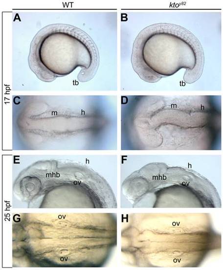
The hindbrain phenotype of ktoy82. Live images of lateral (A,B,E,F) and dorsal views (C, D, G, H) of the developing hindbrain at 17 hpf (A–D) and 25 hpf (E–H). The early midbrain and hindbrain regions are malformed at 17 hpf in mutant embryos (D), and no ventricle is visible at 25 hpf (H). h, hindbrain; m, midbrain; mhb, mid-hindbrain boundary; ov, otic vesicle; tb, tail bud. PHENOTYPE:
|
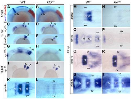
Hindbrain segmentation in the ktoy82 mutant. Lateral (A–J) and dorsal views (K–T) of wt and mutant embryos, as indicated, stained by whole mount in situ hybridization. Two-color in situ hybridization with wnt1 (blue) and hoxd4a (red) at 17 hpf (A,B). Expression of egr2b (C,D) and mafba (E,F) at 19 hpf, and cyp26c1, egr2b, epha4a, mafba, wnt8b, hoxb1a and efnb3b at 25 hpf (G–T). mhb, mid-hindbrain boundary; ov, otic vesicle; r, rhombomere. EXPRESSION / LABELING:
|
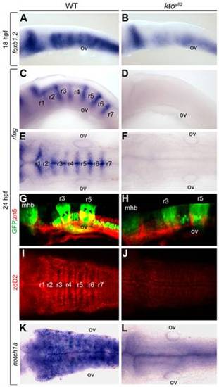
Specific loss of hindbrain boundaries in ktoy82 mutants. Lateral (A–D,G,H) and dorsal views (E,F,I–L) of wt (A,C,E,G,I,K) and ktoy82 mutant embryos (B,D,F,H,J,L) at 18 hpf (A,B) and 24 hpf (C–L). (A–F, K,L) in situ hybridization. (A,B) Expression of the earliest hindbrain boundary marker foxb1.2. (C–F) Completely loss of rfng expression in hindbrain boundaries. (G–J) Confocal images of hindbrain boundary neurons; immunostaining with zn5 (red) in pGFP-5.3 transgenic zebrafish (green) (G,H); staining with zebrafish delta D antibody, zdD2 (I,J). (K,L) notch1a expression in the hindbrain. mhb, mid-hindbrain boundary; ov, otic vesicle; r, rhombomere. EXPRESSION / LABELING:
PHENOTYPE:
|
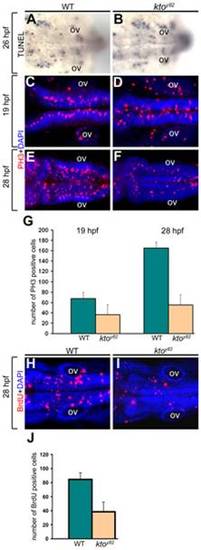
Cell proliferation defects in the hindbrain of ktoy82 mutants. All images are dorsal views at 19 hpf (C,D), 26 hpf (A,B), and 28 hpf (E,F,H,I). (A,B) Analysis of cell death using TUNEL assay. (C–G) PH3 staining (C–F), and quantification (G) of PH3 positive cells in wt and kto embryos. (H–J) BrdU staining of wt and mutant embryos (H,I), and quantification of BrdU positive cells (J). Five embryos were counted for each condition; the error bars indicate 1 standard deviation based on 5 samples. PH3 staining at 19 hpf, wt vs. kto: p = 0.003456; at 28 hpf, wt vs. kto: p = 2.97e-05. BrdU staining, wt vs. kto: p = 0.000706. ov, otic vesicle. PHENOTYPE:
|
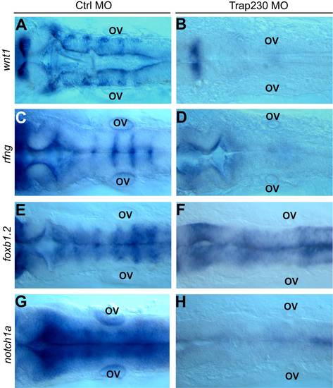
Injection of Med12 MO recapitulates the hindbrain boundary phenotype of the kto mutant. All images are dorsal views of control MO (A,C,E,G) and Med12 MO (formerly called Trap230 MO) (B,D,F,H) injected embryos at 24 hpf. Wnt1 (A,B), rfng (C,D), foxb1.2 (E,F), and notch1a (G,H) were used as hindbrain boundary markers. ov, otic vesicle. |