
- Title
-
Completion of the epithelial to mesenchymal transition in zebrafish mesoderm requires Spadetail
- Authors
- Row, R.H., Maître, J.L., Martin, B.L., Stockinger, P., Heisenberg, C.P., and Kimelman, D.
- Source
- Full text @ Dev. Biol.
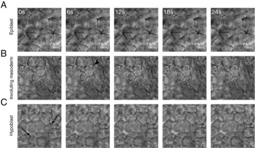
Cell morphology changes during mesoderm maturation. Cell morphology was observed using time-lapse DIC microscopy of the lateral margin during early gastrulation. (A) Cells in the outer (epiblast) layer did not extend protrusions and maintained long, stable, edge-to-edge contacts with their neighbors. (B) As cells underwent involution at the margin they remained tightly packed but extended many protrusions, primarily blebs (arrowhead). (C) Cells in the hypoblast displayed a mesenchymal morphology. They extended actin-based protrusions (arrows) as well as blebs, did not pack tightly, and formed only transient adhesions at discrete points of contact. All images are at the same magnification, scale bar is 10 μM. |
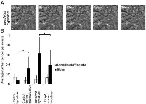
Mesoderm in spt morphants displays abnormal morphology. (A) Cells in the hypoblast of spt morphant embryos blebbed extensively and failed to adopt the proper mesenchymal morphology. (B) Cell protrusions were quantified in wild-type embryos during and after involution, in the hypoblast of spt morphants and in spt morphants expressing spt under the control of a heat shock-sensitive promoter. Mesodermal cells in spt morphants blebbed far more than in wild-type hypoblast, whereas restoring Spadetail significantly reduced this blebbing. Error bars indicate one standard deviation. At least 22 cells were quantified per condition. p < 0.05 for data marked with an asterisk. |
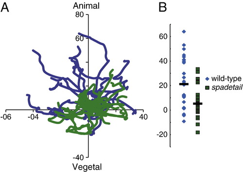
spt morphant mesoderm migrates abnormally. (A) Recently involuted cells were tracked for 30 min in wild-type (blue traces) and spt morphant (green traces) embryos at 55-60% epiboly, lateral to the shield. The x-axis is aligned with the margin of the embryo. spt morphant cells failed to exhibit the animal-ward bias of migration observed in wild-type mesoderm. A representative set of tracks from one wild-type and one spt morphant is shown. (B) The final positions of all tracked cells on the y-axis are marked, with bars indicating median positions. Wild-type cells had a significant migration bias towards the animal pole, much greater than was observed in spt morphant cells. All axes are labeled with µm of displacement from the starting position. These results are taken from tracking 26 cells from 3 embryos, per condition. |
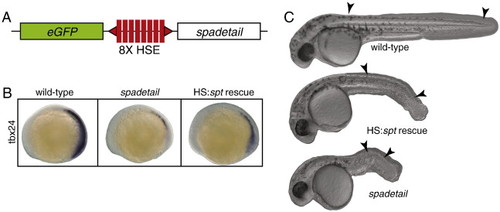
Restoring Spadetail significantly rescues the spt phenotypes. (A) A genetic construct was incorporated into the zebrafish genome by Tol2-mediated transgenesis. The spt and eGFP genes were placed downstream of a bi-directional heat shock-sensitive promoter. (B) Expressing spt from this construct partially restored expression of tbx24 in spt morphant embryos. (C) Significant phenotypic rescue was also observed by inducing transgene expression early in gastrulation. Arrowheads mark the position of the first and last formed somite in each embryo. PHENOTYPE:
|
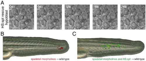
Cell morphology and cell migration are rescued by restoring Spadetail. (A) Cells in the hypoblast of spt morphant cells expressing spt after transgene induction blebbed less frequently than with morpholino alone (see Fig. 2B for quantification). (B) When spt morphant cells were transplanted into the ventral mesoderm of a wild-type embryo at shield stage they were unable to contribute to somites. (C) Restoring Spadetail by inducing transgene expression allowed cells to migrate out of the tailbud and contribute to developing somites. Each panel is representative of at least 16 successful transplants. |
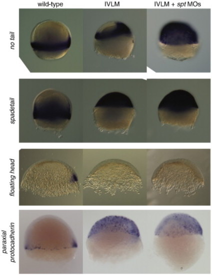
Mesoderm marker gene expression in induced ventral/lateral mesoderm embryos. The utility of the induced ventral/lateral mesoderm system was demonstrated by analyzing marker gene expression in treated embryos. Mesodermal identity was assayed by in situ hybridization for transcripts typical of ventral/lateral mesoderm fate (no tail (ntl), spadetail (spt) and paraxial protocadherin (papc)) or axial mesoderm fate (floating head (flh)). Treated embryos greatly expand their expression of ventral/lateral marker genes while axial fates are repressed. Blocking expression of spt with morpholino injection expands the domain of ntl expression and partially blocks papc expression, similar to what is observed in the mesoderm of spt embryos. The view is lateral with the dorsal pole to the right when it could be identified. |
Reprinted from Developmental Biology, 354(1), Row, R.H., Maître, J.L., Martin, B.L., Stockinger, P., Heisenberg, C.P., and Kimelman, D., Completion of the epithelial to mesenchymal transition in zebrafish mesoderm requires Spadetail, 102-110, Copyright (2011) with permission from Elsevier. Full text @ Dev. Biol.