
- Title
-
Drug screening in a zebrafish model of Duchenne muscular dystrophy
- Authors
- Kawahara, G., Karpf, J.A., Myers, J.A., Alexander, M.S., Guyon, J.R., and Kunkel, L.M.
- Source
- Full text @ Proc. Natl. Acad. Sci. USA
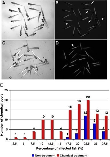
Fish at 4 dpf from screening with the Prestwick library on heterozygous sapje fish pairs and the percentage of affected fish after chemical treatment with these chemical pools. Fish with nontreatment (A and B) and chemical treatment (C and D). (A and C) Bright image. (B and D) Birefringence image. In 20 embryos, five apparently affected fish are caused by a homozygous mutation in the zebrafish dystrophin gene, which is inherited in a recessive manner (25%; arrows). In fish treated with chemical pools, only one fish (5%; arrow) shows the muscle phenotype by birefringence. (E) Distribution of the percentage of affected fish treated with chemical pools and untreated fish. A total of 140 chemical pools (1,120 total chemicals) were tested. Treatment with 108 of the chemical pools was not lethal to the fish. Blue bars, untreated fish (n = 28; independent determination). Red bars, chemically treated fish with 108 chemical pools (in duplicate). Some chemical pools decreased the percentage of affected fish showing abnormal birefringence compared with untreated fish and might contain a compound able to restore normal muscle structure. |
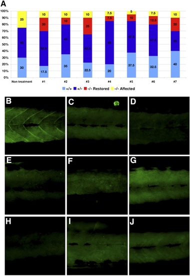
Genotype and dystrophin expression of treated fish with seven candidate chemicals from the longer screen. (A) Genotyping of each of 40 fish treated with each of the seven potential candidate chemicals. Yellow, dystrophin-null affected fish (abnormal birefringence). Red, dystrophin-null unaffected fish (normal birefringence). Blue, heterozygous fish (normal birefringence). Light blue, WT fish (normal birefringence). In each of the seven chemically treated wells, some fish show normal birefringence despite genotyping results that show dystrophin-null fish. (B–J) Immunostaining with an anti-dystrophin antibody. (B) WT. (C) Dystrophin-null fish. (D–J) Dystrophin immunostaining of fish with restored muscle structure with each of the seven chemicals (chemicals 1–7 in Table 2). All restored fish with chemicals 1–7 showed no expression of dystrophin, similar to those of untreated dystrophin-null fish (C). EXPRESSION / LABELING:
PHENOTYPE:
|
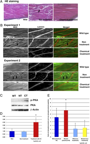
Analysis of skeletal muscle of dystrophin-null fish that survive 30 d after treatment with chemical 4, aminophylline. (A) H&E staining of WT, nontreated, and chemical 4-treated fish. Nontreated dystrophin-null fish have disorganized structure (arrow) in skeletal muscle. The dystrophin-null fish with treated aminophylline have normal structure in skeletal muscle. (B) Immunostaining of WT, nontreated, and chemical 4-treated fish with anti-myosin heavy chain and anti-laminin antibody. Nontreated dystrophin-null fish have broken and disturbed structure (arrows) of skeletal muscle fibers. The treated dystrophin-null fish with aminophylline have normal structure in skeletal muscle. (C) Immunoblot of phosphorylated PKA and PKA at 30 dpf in surviving fish. WT, wild type; NT, nontreatment of dystrophin-null fish; CT, chemical 4-treated dystrophin-null fish. (D) Ratio of phosphorylated PKA and PKA in 30 dpf surviving fish (n = 3). Blue, WT; light blue, nontreatment of dystrophin-null fish; red, chemical 4-treated dystrophin-null fish. *P < 0.05 (vs. WT and nontreatment groups). Error bars indicate SDs. (E) Assay of cAMP-dependent PK activity (n = 3). Bars show the ratio of phosphorylated peptides and nonphosphorylated peptide. Blue, WT fish treated with chemical 4 (2.5 μg/mL) for 30 d. Red, affected sapje fish treated with chemical 4 (2.5 μg/mL). Light blue, nontreated WT fish. Yellow, nontreated affected sapje fish. *P < 0.05 (vs. nontreated WT and mutant fish). Error bars indicate SDs. PHENOTYPE:
|

ZFIN is incorporating published figure images and captions as part of an ongoing project. Figures from some publications have not yet been curated, or are not available for display because of copyright restrictions. PHENOTYPE:
|
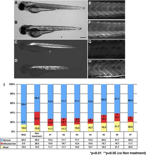
Dystrophin morphant by injection of morpholino and the effects of candidate chemicals in recovering the phenotypes of the morphants. The dystrophin morphant has a similar phenotype to the sapje fish, which shows reduced birefringence and expression of dystrophin. (A, C, E, and F) WT. (B, D, G, and H) Dystrophin morphant. (A and B) Normal light image. (C and D) Birefringence image. (E and G) Immunostaining with anti-dystrophin antibody. (F and H) Immunostaining with anti-laminin antibody. (I) Percentage of dead affected and normal fish in morphants with treatment of each of the seven chemicals. In untreated dystrophin morphants, 30% are affected fish. For each chemical treatment, the percentage of affected fish is reduced compared with those of untreated morphants. Blue bar, normal. Red bar, affected fish. Yellow bar, dead fish (*P < 0.01, **P < 0.05; n = 3). |