
- Title
-
Control of cell migration in the development of the posterior lateral line: antagonistic interactions between the chemokine receptors CXCR4 and CXCR7/RDC1
- Authors
- Dambly-Chaudière, C., Cubedo, N., and Ghysen, A.
- Source
- Full text @ BMC Dev. Biol.
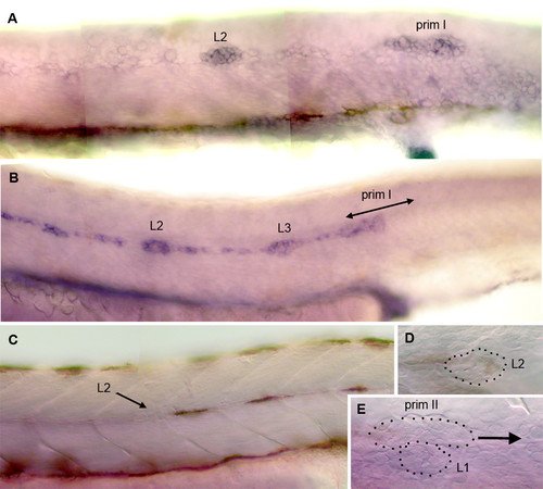
Patterns of cxcr7 expression in the developing PLL. A: expression of claudin [34] reveals the migrating primordium primI and a freshly deposited proneuromast, L2, in a 32 hpf embryo. B: cxcr7 is expressed in the trailing cells of the migrating primordium but not in the leading cells. It is also expressed in proneuromasts L2 and L3 in a 32 hpf embryo, as well as in the trail of interneuromastic cells. C-E: cxcr7 expression has disappeared at 48 hpf, both in the interneuromastic cells and in primary neuromasts. E: cxcr7 is not detectably expressed in the secondary primordium, primII, which is in the process of migrating past L1 at 48 hpf. In all panels the primordium migrates to the right. The dots in D, E outline deposited neuromasts and migrating primordium. |
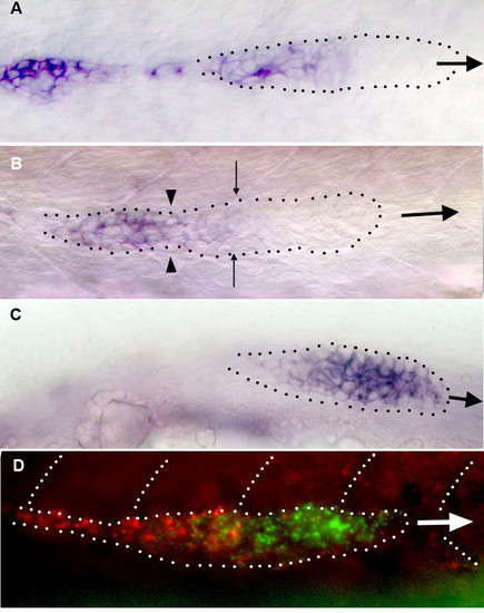
Patterns of gene expression in migrating primordia. A: the expression of cxcr7 is strong in the trailing third of primI and absent in the leading half. B: neuromast deposition depletes the cells that express cxcr7 most strongly (left of the arrowheads) but cells at the trailing edge of the primordium also express cxcr7, albeit at a lower level. The expression of cxcr7 must be quickly up-regulated in these cells to re-establish the pre-deposition pattern (compare B and A). C: the expression of cxcr4 is high in most of the primordium but weaker in the trailing cells. D: a double in situ hybridization reveals that the patterns of expression of cxcr7 (red) and cxcr4b (green) are largely complementary but not exclusive. In all panels the large arrow shows the direction of primordium migration and the dots outline the migrating primordium. In panel B the arrowheads indicate the limit between the group of cells that are slowing down to form a neuromast, and those that will keep migrating. The fine arrows indicate the boundary of the region of cxcr7 expression. EXPRESSION / LABELING:
|
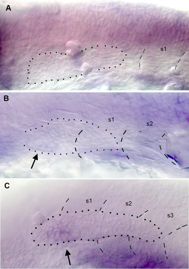
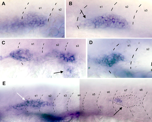
Early expression of cxcr7. A: no expression can be detected in the placode or in the primordium that begins to elongate towards the first somite at 20 hpf. B: expression can first be detected in trailing cells (arrow) when the primordium begins to extend along the myoseptum. C: expression increases (arrow) as the primordium extends along somites 1–3. EXPRESSION / LABELING:
|
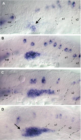
Early expression of cxcr4b. A: expression is first detected in a cluster of placodal cells (arrow) in 19 hpf embryos. B: expression is much enhanced in 20 hpf embryos. C: by 22 hpf the primordium has made contact with the SDF1 trail and elongates into somite 1. D: at about the same time cxcr4 expression is down-regulated in a small cluster of cells near the presumptive trailing edge of the primordium (arrow). EXPRESSION / LABELING:
|
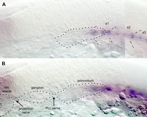
Early expression of sdf1a and primordium migration. A: At 22 hpf a thin stripe of cells along the horizontal myoseptum begins to express sdf1a. Dotted outline of the primordium derived from panel B. B: in the same embryo but in a slightly more superficial focal plane the primordium extends posteriorly and overlays the tip of the sdf1a stripe. EXPRESSION / LABELING:
|
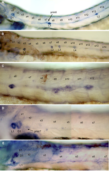
Morphant phenotypes. A: in wild-type 48 hpf embryos, alkaline phosphatase activity is present in the neuromasts, in the trail of interneuromastic cells and more weakly in primII (arrowed). The first neuromast of the dorsal line, D1, is already present at this stage. B: moderate phenotype in cxcr7 morphant embryos: there are fewer neuromasts (in this embryo, 4 instead of 7–8) and they are positioned closer together (see also Fig. 8). C: in about 10% of the cases the primoridum fragments in 2–3 clusters as is normally seen only for the terminal neuromasts at the tip of the tail. D: strong phenotype of a cxcr7 morphant embryo: no migration has taken place and there is a single neuromast, L1, at the level of the first somite. The first neuromast of the dorsal line, D1, has also formed. D1 can be unambiguously identified due to the anisotropy in alkaline phosphatase labeling which is orthogonal to that of the L neuromasts (panel B and [24]). A group of cells that may correspond to either primI or primII (arrowed) is stalled on somite 2. PHENOTYPE:
|
Expression of cxcr4b and cxcr7 in morphant embryos. A, B: expression of cxcr4b in 32hpf Mo-cxcr7 embryos. A: the shape of the stalled primordium is usually round and expression of cxcr4 appears ubiquitous. B: in a primordium that has migrated over a few somites, the expression of cxcr4b is reduced in the trailing cells (arrow), but the expression appears less asymmetric than in wild type embryos (compare with Fig. 3C). C, E: Expression of cxcr7 in 32hpf cxcr4b-MO1 embryos. C: in this embryo part of the primordium has reached somite 2–3 and has begun to extend over the yolk (arrow). All cells express cxcr7. E: an exceptional case where half of the primordium has remained around somite 1 and the other half has migrated up to somite 8 (in a normal embryo the primordium would have reached somite 15–20 at this age). In the stalled group all cells express cxcr7 while in the migrating group only the trailing cells express this gene. D: inactivation of SDF1 leads to the stalling of the primordium and to the expression of cxcr7 in all cells. Dashed lines indicate the positions of somite borders and dots in panel E show the outline of the primordium, as seen under Nomarski optics. |

Unillustrated author statements PHENOTYPE:
|