
- Title
-
Sox17 and chordin are required for formation of Kupffer's vesicle and left-right asymmetry determination in zebrafish
- Authors
- Aamar, E., and Dawid, I.B.
- Source
- Full text @ Dev. Dyn.
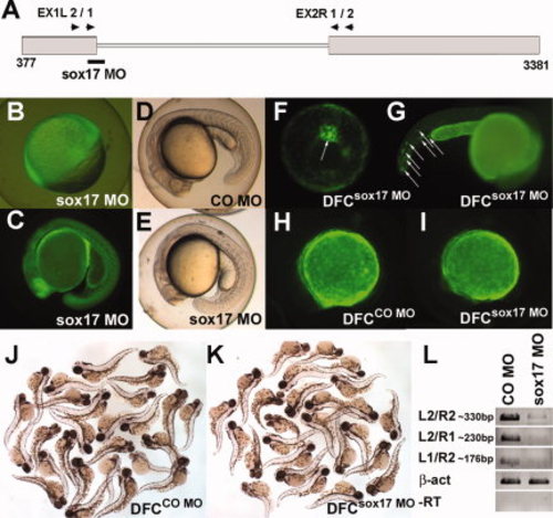
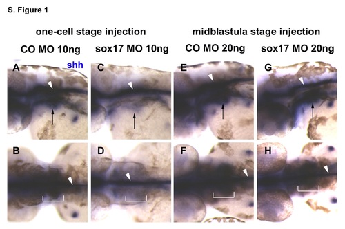
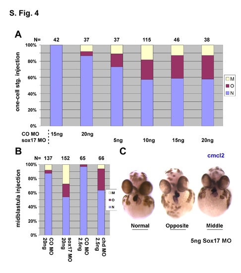
Global phenotypes arise in sox17 morphants, but not in DFCsox17 MO embryos. A: Location of the sox17 MO. 32-Fluorescein tagged sox17 MO was widely distributed in the embryos injected at the one-cell stage (B: shield; C: late somite stage). D,E: 22hpf-old embryos injected at the one-cell stage with 10 ng control (CO) MO, and sox17 MO, respectively. F-I: Injection into the yolk at the midblastula stage excludes the MO from most embryonic cells except the DFC (F, arrow, 80% epiboly) and a few cells in the tail (G, arrows. 1dpf). H–K: Embryos injected into the yolk at the midblastula stage with 20 ng CO MO (H,J) or sox17 MO (I,K) were similar. Pictures taken under fluorescence illumination at about 8-somite stage (H,I), and in visible light at 2dpf (J,K). L: The efficiency of sox17 MO, 10 ng, injected at the one-cell stage, was assessed at 60-80% epiboly by RT-PCR with primers shown in A; β-act was used as control. All pictures are lateral view, except vegetal view with dorsal on top in F. C-E and H-I: anterior to the left; G: anterior to the right. |

Injection of sox17 MO leads to abnormal pancreas placement, which is rescued by sox17 RNA co-injection. Embryos injected at the one-cell stage with 10 ng CO MO, or 10 ng sox17 MO alone or combined with sox17 RNA as indicated, were in situ hybridized with insulin at 3dpf. A-C: Sox17 MO injected embryos showed normal expression on the right side (A), and abnormal opposite expression on the left (B); dorsal views, anterior to left. Some embryos showed fragmented ins-positive domains (C left, arrows), with an example of an intact domain in the right panel (lateral views, anterior to right). D: Localization of ins expression after injection of MOs with or without RNA, as indicated. R, right; M, middle; L, left; f in Rf, Mf, or Lf refers to fragmented ins-positive domains. Number of embryos is shown at the top. EXPRESSION / LABELING:
PHENOTYPE:
|
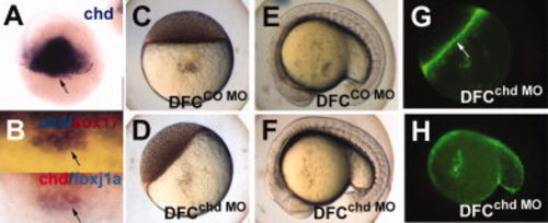
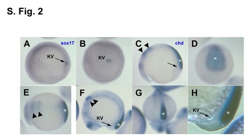
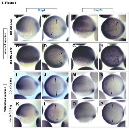
Chordin expression in the dorsal forerunner cells in zebrafish embryos and targeting of DFC by chd MO. A,B: Whole mount in situ hybridization for chd at 50% epiboly (A), and double staining of chd (blue) and sox17 7 (red) (B, top), and chd (red) and foxj1a (blue) (B, bottom) at 60% epiboly; arrows point to DFC. Embryos were injected at the midblastula stage with 2.5 ng CO MO (C,E) or chd MO (D,F), and photographed at the oblong (C,D) and 16-18 somite (E,F) stages. The embryos in D,F are also shown as fluorescent images (G,H, respectively). Arrow in G, yolk syncycial layer. A,B, dorsal views; E,F,H, lateral views, with anterior to the left. EXPRESSION / LABELING:
PHENOTYPE:
|
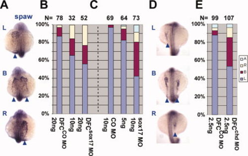
Sox17 and chd are required for left-right asymmetry determination in zebrafish. Embryos were injected into the yolk at one-cell (C) or midblastula (A,B,D,E) stages, with CO MO (B,C,E), sox17 MO (A and B,C), or chd MO (D and E) as indicated in the bar graphs. Whole mount in situ hybridization for spaw (A,D: arrowheads, 15-18 somite; dorsal view with anterior on top) showed aberrant expression when either sox17 MO or chd MO was injected, as summarized in B,C, and E. L, left; B, bilateral; R, right; A, absent. Number of embryos is shown at the top. EXPRESSION / LABELING:
PHENOTYPE:
|
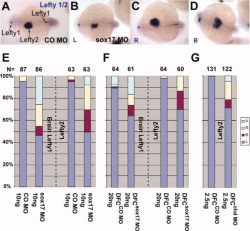
Lefty1,2 expression after suppression of sox17 and of chd in DFC. Embryos were injected into the yolk at one-cell (A–E) or midblastula (F,G) stages, with CO MO (A and E-G), sox17 MO (B-D and E,F), or chd MO (G) as indicated. Whole mount in situ hybridization for lefty1 and/or lefty2 at 22hpf (A-D: arrows; dorsal view with anterior to the left) showed aberrant expression when either sox17 MO or chd MO were injected, as summarized in E-G. Expression of lefty1 in the brain and lefty2 in the heart field was scored, as indicated (E, F) in sox17 MO-injected embryos, but only lefty2 was scored after chd MO injection (G). L, left; B, bilateral; R, right; A, absent. Number of embryos is shown at the top. EXPRESSION / LABELING:
PHENOTYPE:
|
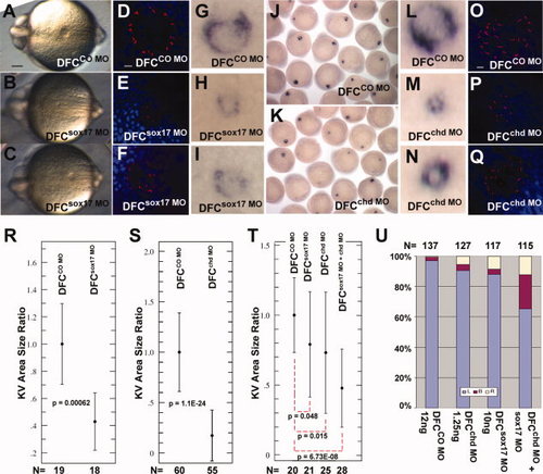
Sox17 and chd are required for proper formation of KV. CO MO (A,D,G: 20 ng; R: 15 ng; J,L,O,S: 2.5 ng; T,U: 12 ng), sox17 MO (B,C,E,F,H,I: 20 ng; R: 15 ng; T,U: 10 ng) or chd MO (K,M,N,P,Q,S: 2.5 ng; T,U: 1.25 ng) were injected into the yolk at the midblastula stage, and embryos were examined at 6–8 somites. Sox17 MO- and chd MO-injected embryos developed small and abnormal KVs as seen in live embryos (A-C; posterior view), by whole mount in situ hybridization with charon (G-N), and by immunostaining for cilia using anti-acetylated tubulin antibody (red) and DAPI (blue) (D-F and O-Q). Scale bars = 0.1 mm in A; 10 µm in D and O. R,S: NIH ImageJ was used to measure the KV area in injected embryos, shown as ratio to the mean control area; error bars are standard deviations; P values are given in the panels. DFCsox17 MO (R) and DFCchd MO (S) embryos showed a significant reduction in KV size compared to controls. T,U: Embryos injected at the midblastula stage with low levels of individual MO (12 ng CO MO, 1.25 ng chd MO, 10 ng sox17 MO) or a combination of 1.25 ng chd MO and 10 ng sox17 MO. KV area was measured as above (T), or lefty2 expression was tested at the -21 somite stage (U). PHENOTYPE:
|
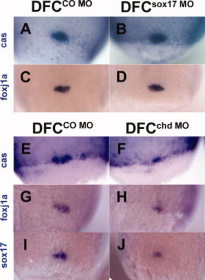
DFC formation in embryos after targeted injection of sox17 MO or chd MO. In situ staining of cas (A,B,E,F), foxj1a (C,D,G,H), and sox17 (I,J) at the 80-90% epiboly stage after midblastula injection of 20 ng sox17 MO (B,D), 20 ng CO MO (A,C), 2.5 ng chd MO (F,H,J), or 2.5 ng CO MO (E,G,I). B and D show no reduction in DFC size whereas F, H, and J show size reduction; sox17 expression also appears reduced (J). All views are dorsal. EXPRESSION / LABELING:
PHENOTYPE:
|
|
|
|
|