
- Title
-
Quantification of vestibular-induced eye movements in zebrafish larvae
- Authors
- Mo, W., Chen, F., Nechiporuk, A., and Nicolson, T.
- Source
- Full text @ BMC Neurosci.
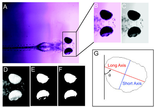
Defining the eye regions in fish larvae. Six steps (A-G) were programmed in MATLAB to quantify eye movements/eye changes in the videos. A head region image (B) was first outlined and extracted from the original image frame (A). The head region was converted into a grayscale image (C) with inverted color (D). Then, the inverted grayscale image was converted into a black-white image (E) using an arbitrary threshold. This black-white image was simplified by removing extra punctae around the retina to define the eye more clearly (F). The final step was to calculate the parameters used to quantify changes during eye movements. (G) Features calculated from the extracted eye region. θ designates the rotation angle. The red line indicates the long axis, and the blue line the short axis of the eye. |
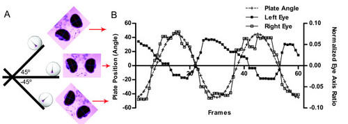
Representative eye movements in response to platform movements (5 dpf). (A) Illustration of the relationship between platform position, larval direction and eye movements. (B) The ratio of long and short axis of both eyes changed sinusoidally. The counter movements of the two eyes followed the platform movements (dashed line). Red arrows between (A) and (B) shows the corresponding position of the larva with respect to platform angle. |

ZFIN is incorporating published figure images and captions as part of an ongoing project. Figures from some publications have not yet been curated, or are not available for display because of copyright restrictions. PHENOTYPE:
|
|
ZFIN is incorporating published figure images and captions as part of an ongoing project. Figures from some publications have not yet been curated, or are not available for display because of copyright restrictions. PHENOTYPE:
|
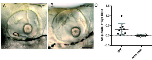
Vestibular-induced eye movements were absent in larvae lacking anterior/utricular otoliths. Representative DIC images depicting lateral views of the inner ear of wild-type (A) and rock solo mutant larvae (B) at 5 dpf. The DIC images were oriented with anterior on the left and posterior on the right. Note the loss of anterior otolith in the mutant, whereas the posterior otolith is unaffected. Scale bar, 100 μm. (C) Amplitudes of eye movements of wild-type siblings and rock solo mutants. PHENOTYPE:
|