
- Title
-
The relationship between dlx and gad1 expression indicates highly conserved genetic pathways in the zebrafish forebrain
- Authors
- Macdonald, R.B., Debiais-Thibaud, M., Talbot, J.C., and Ekker, M.
- Source
- Full text @ Dev. Dyn.
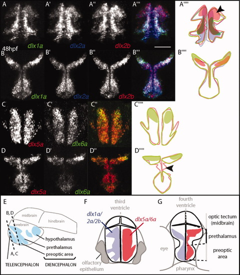
Similar forebrain expression of the two genes from a dlx cluster. A-D″: Single z transverse sections of triple fluorescent in situ hybridization shows dlx1a, dlx2a, and dlx2b expression domains at 48 hours postfertilization (hpf) in the telencephalon (A-A′″) and diencephalon (B,B′″), as well as dlx5a and dlx6a (C,C′″ and D-D′″). Schematic summaries representing the overlaps in dlx expression are shown in A″″, B″″, C″″, and D″″; the solid lines define the boundaries of detectable expression; the zones of relatively higher expression are colored when applicable. The boundaries of expression domains are comparable for dlx1a, dlx2a, and dlx2b, but dlx2b specifically displays a more intense zone of expression in a dorsal-lateral domain (arrow in A″″). Boundaries of expression domains for dlx5a and dlx6a are overall similar although dlx6a was hardly detected in the ventral domain of the prethalamus (arrowhead on D″″). E: Levels of section are shown on the schematic. Shared expression domains of clustered genes (and their paralogs) are mapped on a schematic representation of a brain section at the level of the telencephalon (F) and of the diencephalon (G): dlx1a/2a/2b in blue and dlx5a/6a in red. Scale bar = 100 μm. EXPRESSION / LABELING:
|
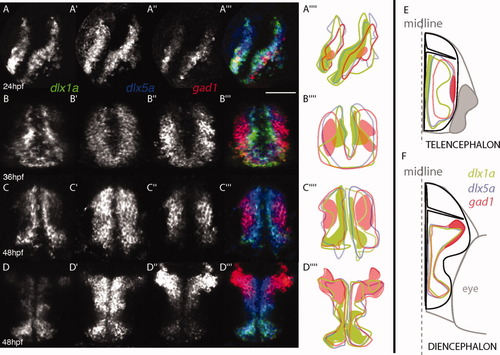
Expression domains of dlx genes with respect to gad1 expression. A-D″: Single z sections of triple fluorescent in situ hybridization for dlx1a, dlx5a, and gad1 in the telencephalon at 24 hours postfertilization (hpf; A,A″, para-sagittal section, anterior is on the left), 36 hpf (B,B″, transverse section of the telencephalon, dorsal is to the top), and 48 hpf (C,C″, transverse section of the telencephalon; D,D″, transverse section of the diencephalon; dorsal is to the top) with a colored merge of all three channels on A′″, B′″, C′″, and D′″. For each section level, a schematic representation is given to localize the boundaries of each expression domain (solid line) and domains of higher levels of expression is represented as a colored surface. E: A schematic representation of the partially nested expression domains in the telencephalon (left half) and diencephalon (right half) at 48 hpf, based on the data shown in C,C′″ and D,D′″: the proximal (ventricular) domain where only dlx1a is detected is shown in green and the lateral domain where gad1 only is detected is colored red. Scale bar = 100 μm. |
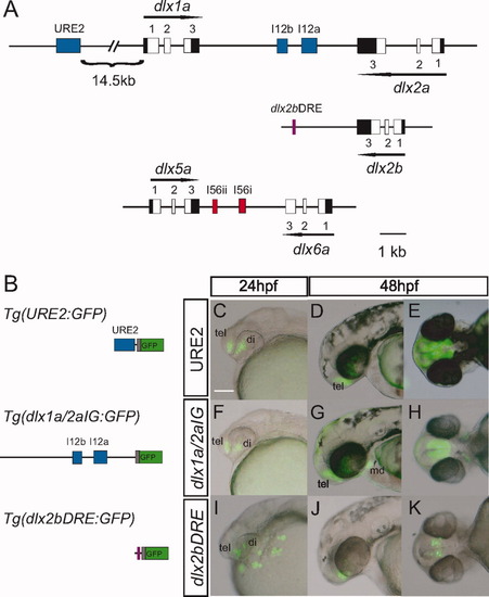
Regulatory elements of dlx1a/2a and dlx2b drive reporter gene expression in the zebrafish forebrain and branchial arches. A: Genomic organization of the dlx1a/dlx2a and of the dlx2b loci. Exons are represented in white and untranslated regions in black. Intergenic regulatory elements are shown as blue (URE2 and I12b) and red boxes (I56i and I56ii). The dlx2bDRE is shown as a purple box. B: Schematic representation of the transgene constructs (containing the β-globin minimal promoter [gray] linked to GFP [green]) used to generate transgenic zebrafish. The Tg(dlx1a/2aIG:GFP) construct contains a 6 kb of the intergenic fragment from the dlx1a/dlx2a intergenic region, the Tg(URE2dlx1a/2a:GFP) contains a 900-bp fragment encompassing the URE2 enhancer and the Tg(dlx2bDRE:GFP) contains a 200-base pair fragment downstream of dlx2b. C-K: Activity of the transgene constructs in the forebrain at 24 hours postfertilization (hpf) and 48 hpf Tg(URE2dlx1a/2a:GFP) transgene (C-E), Tg(dlx1a/2aIG:GFP) transgene (F-H), and Tg(dlx2bDRE:GFP) (I-K). tel, telencephalon; di, diencephalon; md, mandibular portion of branchial arches; GFP, green fluorescent protein. Scale bar = 100 μm. |

The dlx regulatory elements recapitulate endogenous dlx expression in the forebrain of the zebrafish at 48 hours postfertilization (hpf). A-F: Whole-mount in situ with probes against dlx1a (A,B), dlx2a (C,D), and gfp (E,F). E,F: Tg(dlx1a/2aIG:GFP) is expressed in the telencephalon, prethalamus, and hypothalamus. There is also expression in the pharyngeal arches (arrowhead), which recapitulates a small portion of the endogenous dlx arch expression domain. G,H: URE2 activity closely resembles the expression of Tg(dlx1a/2aIG:GFP) in the telencephalon, prethalamus, and hypothalamus and mimics dlx expression in these tissues. tel, telencephalon; pTh, prethalamus; hyp, hypothalamus; di, diencephalon; md, mandible; ba, branchial arches; GFP, green fluorescent protein. Scale bar = 100 μm. EXPRESSION / LABELING:
|
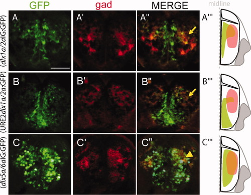
Immunolocalization of green fluorescent protein (GFP) and Gad proteins on transverse cryosections of the telencephalon at 48 hours postfertilization (hpf). A-C″: Expression of GFP (A,B,C) and Gad proteins (A′,B′,C′) is detected in the three transgenic lines, a merged image is presented in A″, B″, and C″ to show overlap (yellow arrows). An interpretative schematic representation of a hemi-section is presented in each case in A′″, B′″, and C′″. Scale bars = 100 μm. EXPRESSION / LABELING:
|
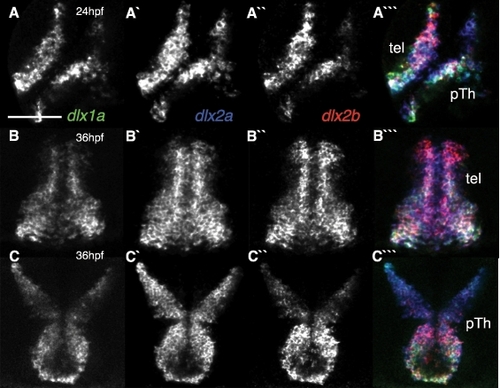
The dlx1a, dlx2a, dlx2b show similar expression in the forebrain at 24 and 36 hours postfertilization (hpf). tel, telencephalon; po, preoptic area; pTh, prethalamus. Scale bar = 100 μm. |
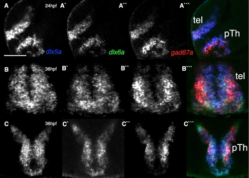
The expression of dlx5a, dlx6a, and gad1 overlaps in the forebrain at 24 and 48 hr postfertilization (hpf). B′″: Expression of dlx5a and dlx6a share overlapping domains with gad1 expression on the lateral boundary. C″: gad1 expression is found throughout the ventral thalamus at 36 hpf. tel, telencephalon; po, preoptic area; pTh, prethalamus. Scale bar = 100 μm. |