
- Title
-
SHIP2, a factor associated with diet-induced obesity and insulin sensitivity, attenuates FGF signaling in vivo
- Authors
- Jurynec, M.J., and Grunwald, D.J.
- Source
- Full text @ Dis. Model. Mech.
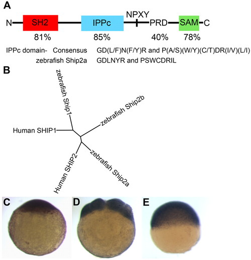
The zebrafish Ship2a protein and expression of maternal ship2a transcripts. (A) The structure of the zebrafish Ship2a protein and the percentage identity between the signature domains of the zebrafish and human SHIP2 protein sequences. The zebrafish Ship2a protein contains motifs that are highly conserved among IPPc catalytic domains. SH2, src homology 2; IPPc, inositol polyphosphate 5-phosphatase catalytic domain; PRD, proline-rich domain; SAM, sterile alpha-motif. (B) Unrooted phylogenetic tree based on Clustal W protein sequence alignments, indicating the relatedness of zebrafish and human SHIP1 and SHIP2 proteins. (C-E) Zebrafish ship2a is maternally supplied and broadly expressed throughout early development. ship2a expression is shown in whole embryos (diameter -0.8 mm) at the one-cell (C), eight-cell (D) and 50% epiboly (5.3 hpf) (E) stages. EXPRESSION / LABELING:
|
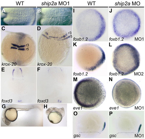
Knockdown of maternal Ship2a dorsalizes the embryo and affects patterning prior to the onset of gastrulation. (A-F) Lateral expansion of dorsal tissue fates in 5–6-somite stage embryos depleted for Ship2a protein. Maternal ship2a-morphant embryos have a lateral expansion of somites (A,B) and a lateral expansion of krox-20 expression in rhombomeres 3 and 5 (r3 and r5) (C,D) compared with WT controls. (E,F) foxd3 expression at the lateral border of the neural plate is reduced in maternal ship2a-morphant embryos. (G,H) ship2a morphants that survive to 24 hpf appear dorsalized. (I–N) foxb1.2 and eve1 expression at the sphere stage (4 hpf) in WT and maternal ship2a-morphant embryos. (I,K) foxb1.2 is expressed dorsally in WT embryos, and (J,L) its expression is expanded to the ventral side of the embryo in maternal ship2a morphants. (M) eve1 expression is restricted to the ventral side in WT embryos, and (N) ventral eve1 expression is lost in maternal ship2a morphants. (O,P) goosecoid (gsc) expression marking the dorsal organizer at 70% epiboly is unchanged between WT and maternal ship2a-morphant embryos. A and B, dorsal views, anterior to the left; C-F, dorsal views, anterior to top; G and H, lateral views; I-N, animal pole views with dorsal to the right; O and P, lateral views, dorsal to right, markings indicate blastoderm margin. EXPRESSION / LABELING:
|
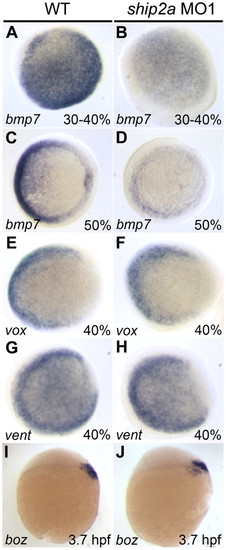
Knockdown of maternal Ship2a alters BMP but not WNT signaling pathways. (A-D) Expression of bmp7 is reduced in 30-40% epiboly and 50% epiboly maternal ship2a morphants as compared with WT controls. (E-J) Gene expression that is dependent on the WNT signaling pathway is not affected in maternal ship2a-morphant embryos. Expression of vox (E,F) or vent (G,H) in 40% epiboly embryos, or bozozok (boz) (I-J) in oblong stage (3.7 hpf) embryos, is unchanged by depletion of maternal Ship2a. A–H, animal pole views with dorsal to the right; I and J, lateral views with animal pole up and dorsal to the right. EXPRESSION / LABELING:
|
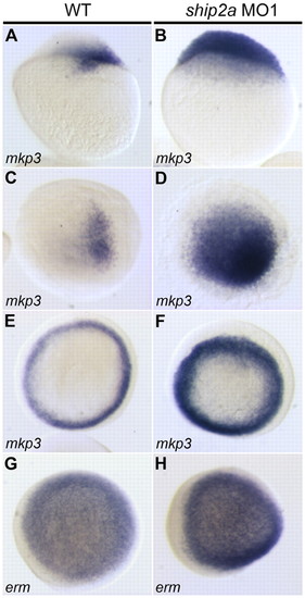
Knockdown of maternal Ship2a affects the expression of FGF-dependent genes in the pre-gastrula embryo. (A,C) mkp3 is expressed dorsally in WT embryos at the oblong stage (3.7 hpf). (B,D) Knockdown of maternal Ship2a results in ventralward expansion of the mkp3 expression domain. (E,F) Expansion of the mkp3 expression domain persists at 50% epiboly (5.3 hpf) in maternal ship2a morphants. (G,H) Expression of erm, a direct target of the FGF signaling pathway, is increased in maternal ship2a morphants. A and B, lateral views with dorsal to the right; C-H, animal pole views with dorsal to the right. EXPRESSION / LABELING:
|
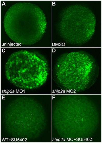
Maternal Ship2a attenuates FGF receptor-mediated pMAPK activation in the pre-gastrula embryo. (A-F) Anti-pMAPK immunoreactivity in oblong-sphere stage (3.7-4 hpf) WT, DMSO-treated control, maternal ship2a morphant and SU5402-treated embryos. (A,B) In WT and DMSO-treated control embryos, pMAPK is restricted to the dorsal side of the embryo. (C,D) Knockdown of maternal Ship2a causes a dramatic ventralward expansion of the pMAPK expression domain. (E,F) Inhibition of FGFR activity with 60 μM SU5402 completely inhibits pMAPK activation in both WT and maternal ship2a-morphant embryos. Confocal projections. Animal pole views with dorsal to the right. EXPRESSION / LABELING:
|
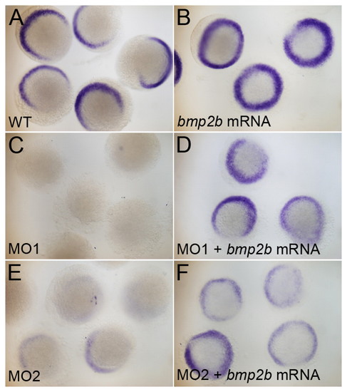
Overexpression of bmp2b restores DV patterning in embryos depleted of maternal Ship2a. (A-F) eve1 expression in WT or ship2a-morphant 50% epiboly embryos and similar embryos that had been injected with bmp2b mRNA. (A,C,E) eve1 expression is restricted to the ventral side of WT embryos and downregulated in ship2a MO1- or MO2-injected embryos. (B) Overexpression of bmp2b in WT embryos results in the dorsalward expansion and circumferential expression of eve1, indicating that ectopic bmp2b expression promotes ventralization. (D,F) Co-injection of bmp2b mRNA with either ship2a MO1 or MO2 restores the WT expression pattern of eve1. |
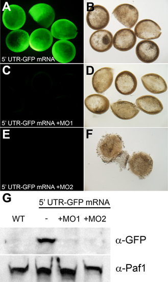
Ability of ship2a MOs to inhibit translation in vivo. (A-F) Expression of GFP in 50% epiboly embryos that were injected at the one-cell stage with 52 UTR GFP mRNA. Embryos were injected with mRNA only (A,B), with mRNA and ship2a MO1 (C,D), or with mRNA and ship2a MO2 (E,F). GFP was detected by immunofluorescence staining. (A,C,E) Fluorescence images (identical exposures times) showing GFP immunostaining and (B,D,F) corresponding fields of embryos under bright field illumination. (G) Immunoblot to detect GFP expression relative to control protein Paf-1 expression in extracts of 50% epiboly embryos that were: not injected (WT), injected at the one-cell stage with 52 UTR GFP mRNA only (-), or injected at the one-cell stage with 52 UTR GFP mRNA along with ship2a MO1 (+MO1) or ship2a MO2 (+MO2). |
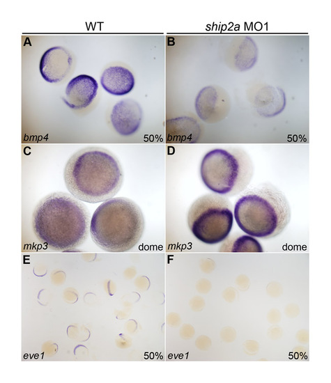
Consistent perturbations of gene expression patterns result from MO-induced depletion of Ship2a protein in zebrafish embryos. Representative groups of WT (A,C,E) or Ship2a-depleted (B,D,F) pre-gastrula embryos. Within each group, stage-specific gene expression patterns are highly reproducible. bmp4 and eve1, genes that are expressed at higher levels on the ventral side of the WT embryo, are downregulated in ship2a morphants. mkp3, a dorsal-specific marker in WT embryos, is expressed throughout the blastoderm margin in ship2a morphants. |