- Title
-
Expression of strawberry notch family genes during zebrafish embryogenesis
- Authors
- Takano, A., Zochi, R., Hibi, M., Terashima, T., and Katsuyama, Y.
- Source
- Full text @ Dev. Dyn.
|
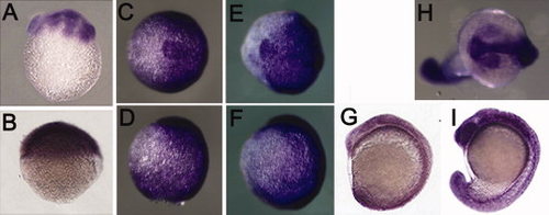
Expression pattern of sbno1 during zebrafish embryogenesis detected by whole-mount in situ hybridization. A: A lateral view of early cleavage stage (2 hours postfertilization [hpf]) embryo. B: A lateral view of 50% epiboly stage (5 hpf) embryo. C,D: A 90% epiboly stage (9 hpf) embryo. E,F: A bud stage (10 hpf) embryo. G,H: A 3-somite stage (11 hpf) embryo. I: A 10-somite stage (15 hpf) embryo. J,K: A 18-somite (18 hpf) stage embryo. C,E,G,J are views from animal pole of the in situ specimen. In D,F,H,I,K, lateral views of in situ specimen is shown with ventral to the left. EXPRESSION / LABELING:
|
|
The embryonic expression pattern of sbno2a is similar to that of sbno1. A: A lateral view of early cleavage stage (1 hour postfertilization [hpf]) embryo. B: A lateral view of 50% epiboly stage (5 hpf) embryo. C,D: A 90% epiboly stage (9 hpf) embryo. E,F: A bud stage (10 hpf) embryo. G: An eight-somite stage embryo. H,I: An 18-somite (18 hpf) stage embryo. C,E,G, and I are views from animal pole of the embryo. In D, F, G, and I, lateral views of the embryo showing ventral to the left. EXPRESSION / LABELING:
|
|
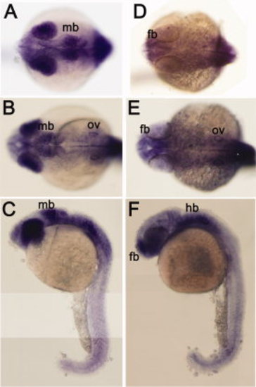
Comparison of expression pattern of sbno1 and sbno2a at pharyngula (24 hours postfertilization [hpf]) stage. A-C: Strong expression of sbno1 was observed in the midbrain (presumptive tectal region) and the eyes at this stage. D-F: Expression of sbno2a is relatively high in the forebrain region at this stage. A and D are focusing on the forebrain region. B and E focus on the hindbrain region. C and F are lateral views of embryos with ventral to the left. fb, forebrain; hb, hindbrain; mb, midbrain; ov, otic vesicle. EXPRESSION / LABELING:
|
|
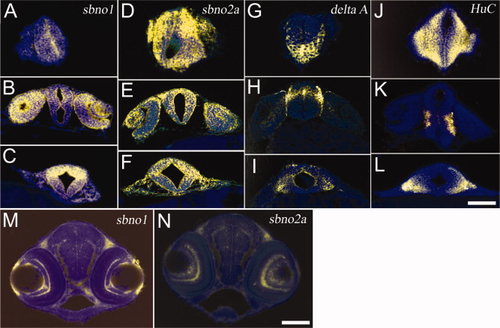
Serial sections of the whole-mount in situ hybridization specimens. A-L: The coronal sections of 24 hours postfertilization (hpf) embryos hybridized to sbno1 (A-C), sbno2a (D-F), delta A (G-I), or HuC (J-L) probes. A,D,G,J: The section 20 to 40 micrometers in depth from the anterior top of the embryos. B,E,H,K: The sections at the anterior midbrain level. C,F,I,L: The sections at the hindbrain level. M,N: The coronal sections of 3 days postfertilization (dpf) larvae at midbrain level hybridized to sbno1 (M) or sbno2a (N) probes. Whole-mount in situ hybridization specimens were embedded in the plastic resin and cut at 10-micrometer thickness. The sections were counterstained by 4′,6-diamidine-2-phenylidole-dihydrochloride (DAPI), and the bright field image showing in situ staining (yellow) and fluorescent image showing DAPI staining (blue) of the same sections were merged. Scale bars = 0.1 mm. EXPRESSION / LABELING:
|
|
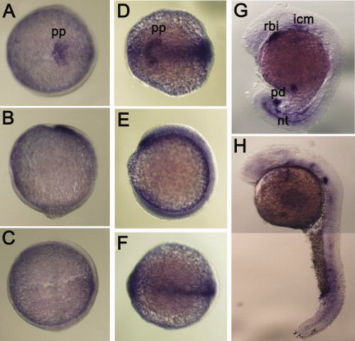
Embryonic expression pattern of sbno2b. A-C: A 90% epiboly stage (9 hpf) embryo. D-F: A bud stage (10 hpf) embryo. G: A 18-somite (18 hpf) stage embryo. H: A pharyngula (24 hpf) stage embryo. A and D are views from animal pole of the embryos. B, E, G, and H are lateral views with ventral to the left. C and F are views from the vegetal pole. Expression of sbno2b was detected in non-neural tissues of embryos, which is distinct from that of the other zebrafish sbno genes. icm, intermediate cell mass; nt, notochord; pd, proctodeum; pp, prepolster; rbi, rostral blood island. EXPRESSION / LABELING:
|
|
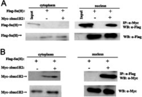
Binding of Su(H)A and sbno1 proteins of zebrafish examined by co-immunoprecipitation assay. A: The protein complex was precipitated using anti-Myc antibody, and the precipitate was detected by Western blot analysis using anti-Flag antibody. B: The protein complex was precipitated using anti-Flag antibody, and the precipitate was detected using anti-Myc antibody. |


