
- Title
-
Functional conservation of a forebrain enhancer from the elephant shark (Callorhinchus milii) in zebrafish and mice
- Authors
- MacDonald, R.B., Debiais-Thibaud, M., Martin, K., Poitras, L., Tay, B.H., Venkatesh, B., and Ekker, M.
- Source
- Full text @ BMC Evol. Biol.
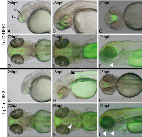
Expression of URE2-GFP reporter transgenes in zebrafish. The Tg-DrURE2 drives GFP expression in the forebrain starting at 24 hpf until at least 96 hpf (A-F). At 96 hpf GFP expression is also observed in the pharyngeal arches (E, F). The Tg-CmURE2 drives GFP expression in the forebrain at 24 hpf until at least 96 hpf (G-L). GFP expression is also first noticed in the somites and pharyngeal arches at 48 hpf (H) and 96 hpf (K, L), respectively. Panels A, B, F, G, H, L are lateral views, anterior to the left and dorsal to the top; panels C, D, E, I, J and K are ventral views. t: telencephalon; d: diencephalon. White arrowheads indicate pharyngeal arch expression and the black arrowhead indicates somite expression. Scale bar: 250μm. |
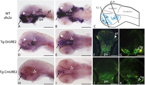
Expression of URE2-GFP reporter constructs in the brain of 48 hpf zebrafish. Expression patterns obtained by in situ hybridization using a dlx2a cDNA probe in wild-type embryos (A, B) or a GFP probe in Tg-DrURE2 (D, E) and in Tg-CmURE2 embryos (H, I). Immunolocalization of GFP proteins on sectioned embryos of the Tg-DrURE2 (F, G) and Tg-CmURE2 (J, K). Expression in the telencephalon is comparable for the endogenous gene and the two transgenes (black arrow in A, B, D, E, H, I). Expression in the dorsal domain of the prethalamus (white arrowhead) in Tg-DrURE2 for gfp mRNA (D, E) and GFP proteins (F) is not observed in Tg-CmURE2 (H-J). Expression of GFP in the hypothalamus (black arrowhead) was restricted to lateral cells in Tg-CmURE2 (H, I, K) compared to Tg-DrURE2 (D, E, G). Panels A, D, H are lateral views, B, E, I are ventral views. Plan for the transversal sections presented in F-G and J-K are localized on the scheme in panel C. Blue domains in the scheme are the forebrain expression domains described for dlx genes: the telencephalic domain being the subpallium (sp, black arrow); the diencephalic domains being the preoptic area (po), prethalamus (pt, white arrowhead) and hypothalamus (hy, black arrowhead). Scale bars: A, B, D, E, H, I, 250 μm; F, G, J, K, 50 μm. EXPRESSION / LABELING:
|
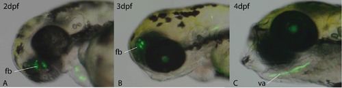
Primary transgenic zebrafish embryos with gfp expressed under MmURE2 sequence. GFP fluorescence could be detected in the forebrain (fb) of primary transgenic zebrafish at 2 dpf (A) and 3 dpf (B) after injection of the construct. GFP fluorescence was also detected in the visceral arches (va) of 4 dpf old embryos after injection (C). |