
- Title
-
Jagged-Notch signaling ensures dorsal skeletal identity in the vertebrate face
- Authors
- Zuniga, E., Stellabotte, F., and Crump, J.G.
- Source
- Full text @ Development

Jag1b-Notch2 signaling regulates DV patterning of the zebrafish facial skeleton. (A,B) Skeletal staining at 5 dpf showing cartilage (blue) and bone (red). jag1bb1105 mutants display a characteristic kink (arrow) behind the eye, which is not seen in the wild type (Wt). (C) Schematic of Jag1b protein showing DSL (blue), EGF-like (white), cysteine-rich (red), transmembrane (TM) and intracellular (yellow) domains. The jag1bb1105 lesion is a nonsense mutation (W223*) that truncates Jag1b in the DSL domain required for Notch binding. (D-G) Dissected facial skeletons from wild-type (D), jag1bb1105 (E), notch2-MO (F) and 20-28 hpf heat shock-treated hsp70I:Gal4; UAS:JAG1 (G) larvae. Schematics (below) show ventral (red) and dorsal (green) elements derived from the mandibular and hyoid arches, with bones more lightly shaded. The maxillary-derived pterygoid process (Ptp) is in gray. Scale bar: 100 µm. (H) Calcein Green bone staining at 5 dpf shows Op-to-Br transformations in jag1bb1105 and notch2-MO larvae and Br-to-Op transformations in 20-28 hpf heat shock-treated hsp70I:Gal4; UAS:JAG1 larvae. (I) The proportion of wild-type, jag1bb1105, jag1b-MO, notch2-MO and 20-28 hpf heat shock-treated hsp70I:Gal4; UAS:JAG1 larvae showing normal (yellow), reduced (red) or transformed (blue) skeletal elements. M, Meckel′s; Pq, palatoquadrate; Hm, hyomandibular; Sy, symplectic; Ch, ceratohyal; Op, opercle bone; Br, branchiostegal ray bone. The proportion of larvae exhibiting ectopic cartilage (blue) is also shown. |
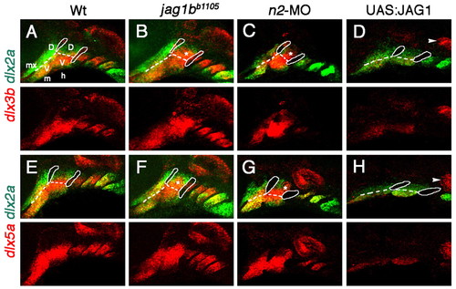
Jag1b-Notch2 signaling inhibits dlx3b and dlx5a expression in the pharyngeal arches. (A-H) Double-fluorescence in situ hybridizations showing the expression of dlx3b or dlx5a (red) and dlx2a (green) at 36 hpf. Compared with wild types (A,E), dlx3b and dlx5a expression is expanded into the dorsal hyoid arches (asterisks) of jag1bb1105 (B,F) and notch2-MO (C,G) zebrafish embryos and is reduced in the ventral arches of 20-28 hpf heat shock-treated hsp70I:Gal4; UAS:JAG1 (D,H) embryos. The expression of dlx3b and dlx5a in otic placodes (arrowheads) is unaffected in UAS:JAG1 embryos. Endodermal pouches (solid lines) and DV arch boundaries (dashed lines) are indicated in the merged images. The maxillary domain (mx) and the dorsal (D) and ventral (V) domains of the mandibular (m) and hyoid (h) arches are indicated for wild type. EXPRESSION / LABELING:
|
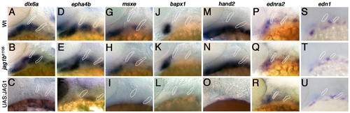
Jag1b generally represses ventral gene expression in the pharyngeal arches. (A-U) In situ hybridizations showing the mandibular and hyoid arch expression of dlx6a (A-C), epha4b (D-F), msxe (G-I), hand2 (M-O), ednra2 (P-R) and edn1 (S-U) at 36 hpf and of bapx1 (J-L) at 40 hpf. The expression of dlx6a, epha4b, msxe and bapx1 is dorsally expanded in jag1bb1105 zebrafish embryos (B,E,H,K) and is greatly reduced in 20-28 hpf heat shock-treated hsp70I:Gal4; UAS:JAG1 embryos (C,F,I,L). hand2 expression is reduced in UAS:JAG1 embryos, whereas ednra2 and edn1 expression is unaffected in jag1bb1105 and UAS:JAG1 embryos. Endodermal pouches are outlined. EXPRESSION / LABELING:
|
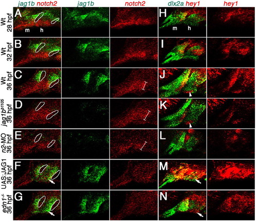
Expression of jag1b, notch2 and hey1 is regulated by Jagged-Notch and Edn1 signaling. (A-N) Confocal sections of double-fluorescence in situ hybridizations showing mandibular (m) and hyoid (h) arch expression of jag1b (green) and notch2 (red) (A-G) and of dlx2a (green) and hey1 (red) (H-N). In wild types, jag1b and hey1 CNCC expression expands ventrally from 28 hpf (A,H) to 32 hpf (B,I) and 36 hpf (C,J). In jag1bb1105 (D,K) and notch2-MO (E,L) zebrafish embryos, jag1b and hey1 expression is reduced and strong ventral notch2 expression (brackets) is expanded at 36 hpf. Arrowheads in J,K indicate ventral mesoderm expression of hey1, which is unaffected in jag1bb1105 mutants. In 20-28 hpf heat shock-treated hsp70I:Gal4; UAS:JAG1 (F,M) and edn1-/- (G,N) embryos, jag1b and hey1 expression is expanded into ventral CNCCs (arrows) and notch2 expression is reduced at 36 hpf. Endodermal pouches are outlined in A-G. |
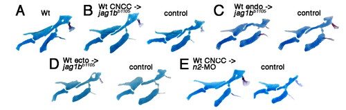
Jag1b-Notch2 signaling functions within CNCCs for DV facial patterning. (A-E) Dissected zebrafish facial skeletons at 5 dpf with wild type (A) shown for reference. Transplantations of wild-type CNCC precursors (B), but not wild-type endoderm (C) or surface ectoderm (D) precursors, rescue the jag1bb1105 skeleton, as compared with the control non-recipient sides. Wild-type CNCC precursor transplants also rescue notch2-MO skeletal defects (E). |
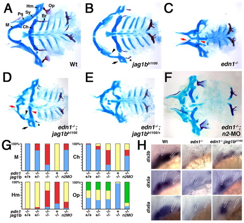
Reduction of Jag1b-Notch2 signaling rescues the ventral defects of edn1 mutants. (A-F) Ventral views of dissected zebrafish facial skeletons at 5 dpf, with elements labeled in wild type (A). jag1bb1105 mutants (B) have Pq reductions (arrowhead) and variable transformations of Hm (arrow) and Op (asterisk). In edn1-/- mutants (C), M (red arrow) and Ch (red arrowhead) are nearly absent. In edn1-/-; jag1bb1105 larvae (D), development of ventral M and Ch is variably restored yet Pq and Hm defects are still evident. M and Ch development is also partially restored in some edn1-/-; jag1bb1105/+ (E) and edn1-/-; notch2-MO (F) larvae. (G) Quantification of skeletal rescue, showing wild-type (yellow), weakly defective (red), severely defective (blue), and expanded (green) cartilage and bone. (H) In situ hybridizations showing dlx3b, dlx5a and dlx6a expression in arch CNCCs at 36 hpf. Compared with edn1-/- embryos, edn1-/-; jag1bb1105 embryos show partial rescue of expression. Endodermal pouches are outlined. See Fig. 1 for abbreviations. |
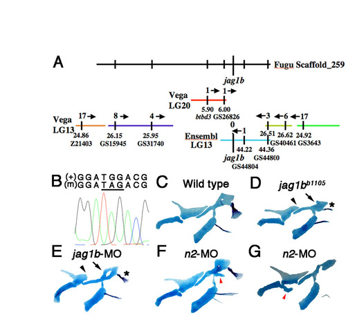
Identification of the jag1bb1105 mutation. (A) Using synteny between Fugu and zebrafish Vega and Ensembl contigs, the b1105 mutation was mapped to a small region of LG13 containing jag1b. Recombinants per 2000 meioses are shown above each contig, and positions in Mb and marker names below. (B) An electrophoretogram showing sequence surrounding the G-to-A transition that creates a premature stop codon (underlined) in jag1bb1105 mutants (m). (C-G) Unilateral flat-mount dissections of 6 dpf facial skeletons stained for cartilage (blue) and bone (red). jag1b-MO larvae show similar skeletal defects to jag1bb1105 mutants, including truncation of Pq (arrowheads), shape changes in Hm (arrows) and transformed Op bone (asterisks). In notch2-MO larvae, ectopic cartilage processes (red arrowheads) are seen at low frequency near the DV interfaces of the hyoid (F) and mandibular (G) skeletons. |
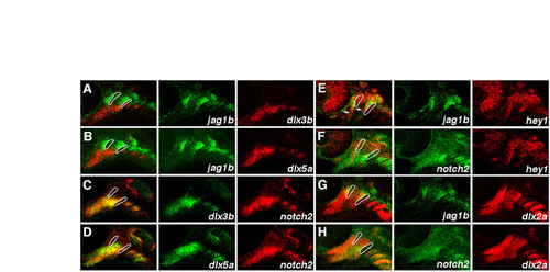
Pharyngeal arch expression of jag1b and notch2 relative to dlx3b, dlx5a, hey1 and dlx2a. (A-H) Double-fluorescence in situ hybridizations at 36 hpf, with endodermal pouch outlines shown for reference in the merged panels. jag1b expression is largely non-overlapping with that of dlx3b and dlx5a. The stronger ventral expression of notch2 overlaps with dlx3b and dlx5a expression, with the border of dlx5a expression extending slightly more dorsally than that of strong notch2 expression. jag1b and hey1 are co-expressed in dorsal CNCCs, with arrows indicating where hey1 expression extends slightly more ventrally than jag1b. hey1 expression also overlaps with weaker dorsal notch2 expression in CNCCs. Colocalization of jag1b and notch2 with the CNCC-specific dlx2a probe confirms that jag1b is expressed in dorsal CNCCs and notch2 is expressed throughout arch CNCCs. |
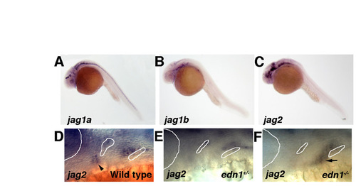
jag2, but not jag1a, is expressed in the pharyngeal arches and is regulated by Edn1. (A-C) Whole-mount in situ immunohistochemistry at 36 hpf. jag1a and jag1b show complementary, non-overlapping expression patterns, with jag1a expression in the central nervous system and jag1b expression in arch CNCCs and sensory and otic placodal cells. jag2 is expressed in the ventral brain and weakly in the pharyngeal arches. (D-F) Higher magnification views of jag2 expression at 36 hpf. In wild types, jag2 expression is restricted to dorsal CNCCs of the mandibular and hyoid arches and the mandibular arch artery (arrowhead). Compared with expression in edn1+/- siblings, jag2 expression in edn1-/- embryos is expanded into the ventral hyoid arch (arrow). Outlines of the eye and endodermal pouches are shown. |
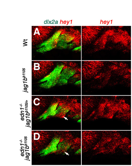
hey1 expression in edn1-/-; jag1bb1105 embryos. (A-D) Double-fluorescence in situ hybridizations showing dlx2a (green) and hey1 (red) expression in the mandibular and hyoid arches at 36 hpf. hey1 is expressed in dorsal CNCCs of wild types (A) and greatly reduced in jag1bb1105 embryos (B). Compared with the ventral expansion seen in edn1-/-; jag1bb1105/+ siblings (C), hey1 expression is reduced but still present at low levels throughout arch CNCCs of edn1-/-; jag1bb1105 double mutants (D). Arrows indicate hey1 expression in ventral CNCCs of edn1-/-; jag1bb1105/+ and edn1-/-; jag1bb1105 embryos. |
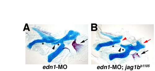
Partial Edn1 reduction does not rescue the dorsal skeletal defects of jag1bb1105 mutants. Flat-mount dissections of 6 dpf facial skeletons stained for cartilage (blue) and bone (red). (A) Wild-type larvae injected with a low dose of edn1-MO display losses of joints (asterisks), a short Sy (black arrowhead) and an expanded Op (black arrow). (B) In jag1bb1105 larvae injected with a low dose of edn1-MO, both edn1-MO and jag1bb1105 phenotypes are seen. Note the transformations of dorsal Hm (red arrow) and Op (black arrow) and the truncation of dorsal Pq (red arrowhead). |