
- Title
-
UDP xylose synthase 1 is required for morphogenesis and histogenesis of the craniofacial skeleton
- Authors
- Eames, B.F., Singer, A., Smith, G.A., Wood, Z.A., Yan, Y.L., He, X., Polizzi, S.J., Catchen, J.M., Rodriguez-Mari, A., Linbo, T., Raible, D.W., and Postlethwait, J.H.
- Source
- Full text @ Dev. Biol.
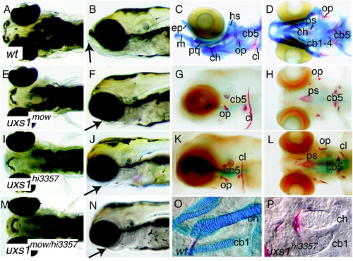
Craniofacial and skeletal phenotypes of zebrafish larvae. Ventral and lateral views of live (A, B, E, F, I, J, M, N) and Alcian blue-, Alizarin red-stained (C, D, G, H, K, L, O, P) animals. Compared to wild types (A, B), mutant animals (E,F moww60 allele; I, J hi3357 allele) had reduced lower jaws (arrows) at 5 dpf. Reduced lower jaw (arrow) in moww60/hi3357 double heterozygotes (M, N) showed failure of complementation. Alcian blue and Alizarin red staining for cartilage (blue) and bone (red) revealed the lack of cartilage and reduced bones in mutants (G, H, K, L) compared to wild type (C, D) at 7 dpf. Nomarski optics on dissected pharyngeal skeletons suggested that mutant cartilages (P) condensed in the same areas as wild types (O), but did not secrete Alcian blue-positive matrix. Abbreviations: cb1-5, ceratobranchials 1 to 5; ch, ceratohyal; cl, cleithrum; ep, ethmoid plate; hs, hyosymplectic; m, Meckel′s cartilage; op, opercle; pq, palatoquadrate; ps, parasphenoid. |
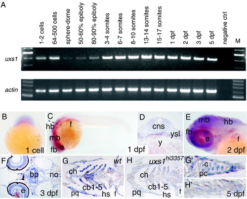
uxs1 expression during zebrafish embryogenesis. (A) RT-PCR for uxs1 transcript in animals of indicated ages, along with β-actin positive controls. Maternal uxs1 mRNA was detected at the 1–2 cell stage and detection decreased at sphere-dome stage. Zygotic uxs1 expression appeared to increase gradually and was maintained at least through 5 dpf. (B) Whole-mount in situ hybridization of a one-cell embryo revealed transcript in the fertilized egg. (C, D) Whole-mount (C) and section (D) of 24 hpf embryos illustrated general expression of uxs1 in brain and craniofacial mesenchyme, as well as in the yolk syncytial layer. The dashed line in panel C indicates the plane of section in panel D. (E) Lateral view of whole-mount 2 dpf embryo showed widespread uxs1 expression in the craniofacial region. (F–H) Horizontal sections of 3 dpf (F) and 5 dpf (G,H) animals. Expression of uxs1 became localized to layers of the retina, brain, and cartilages of the pharyngeal arches. Levels of uxs1 transcript were severely reduced or absent in pharyngeal regions of uxs1hi3357 embryos. High magnification of 5 dpf ceratohyals shows uxs1 expression in both chondrocytes (c) and perichondral cells (pc) of wild types (G′), but low transcript levels in uxs1hi3357 embryos (H′). Abbreviations: bp, basal plate; c, chondrocyte; cb1-5, ceratobranchials 1-5; ch, ceratohyal; e, eye; f, fin bud; fb, forebrain; hb, hindbrain; hs, hyosymplectic; mb, midbrain; no, notochord; pc, perichondrium; pq, palatoquadrate; y, yolk; ysl, yolk syncytial layer. |
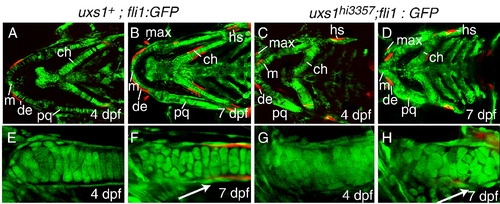
Cellular visualization of cartilage and bone morphologies in wild-type and uxs1 mutant larvae. (A–H) Optical sections of live Alizarin red-stained Tg(fli1:EGFP)y1 larvae, ventral views, at 4 dpf and the same individuals at 7 dpf. (E–H) Focus on the ceratohyal. In wild types (A, B, E, F), chondrocytes stacked and were lined with a flattened layer of perichondral cells (white arrow in F). Ossification centers stained with Alizarin red, reflecting perichondral bone formation in the ceratohyal and hyosymplectic and intramembranous ossification in the dentary and maxilla. In homozygous uxs1hi3357 animals (C, D, G, H); however, chondrocytes were disorganized, the perichondral sheath did not align properly (white arrow in H), and Alizarin red-positive ossification centers (dentary, maxilla, and ceratohyal) were severely reduced in perichondral and intramembranous sites. Abbreviations: ch, ceratohyal; de, dentary; hs, hyosymplectic; m, Meckel′s cartilage; max, maxilla; pq, palatoquadrate. |
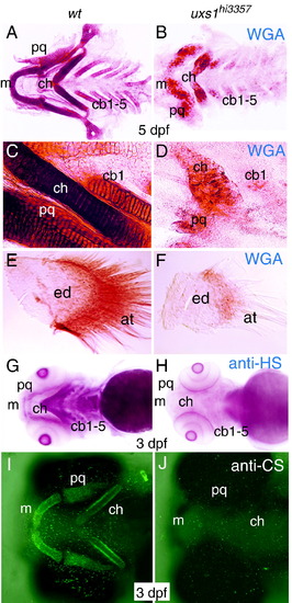
Proteoglycan detection in wild-type and uxs1 mutant skeletons. (A–F) Whole-mount wheat germ agglutinin (WGA) staining to visualize N-acetylglucosamine, ventral views. (G–J) Whole-mount immunostaining against heparan sulfate (G, H) and chondroitin sulfate (I, J) proteoglycans, ventral views. Dissected pharyngeal cartilages revealed reduced WGA staining in uxs1hi3357 mutants (B, D), compared to wild-type siblings (A, C) at 5 dpf. Higher magnification of ceratohyal regions also showed that WGA-positive material was not deposited normally in mutants (D), compared to organized deposition in wild types (C). Dissected pectoral fins showed that both endoskeletal disc and actinotrichia had less WGA staining and fewer actinotrichia in uxs1hi3357 mutants (F), compared to wild-type siblings (E) at 5 dpf. Immunodetection of heparan sulfate demonstrated that HSPGs were localized to pharyngeal domains in wild type (G), but HSPGs were not detectable in homozygous uxs1hi3357 animals (H). Similarly, immunodetection of chondroitin sulfate was abundant in wild-type cartilages (I), but was absent in uxs1 mutants (J). Abbreviations: at, actinotrichia; cb1-5, ceratobranchials 1-5; ch, ceratohyal; ed, endoskeletal disc; m, Meckel′s cartilage; pq, palatoquadrate. EXPRESSION / LABELING:
PHENOTYPE:
|
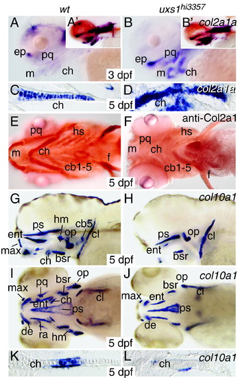
Collagen detection in wild-type and uxs1 mutant skeletons. (A–D) Whole-mount (A, B) and horizontal section (C, D) in situ hybridization for col2a1a gene expression; (E, F) whole-mount immunostaining for Col2 protein; (G–L) whole-mount (G–J) and horizontal section (K, L) in situ hybridization for col10a1 gene expression. Expression of col2a1a increased in developing cartilage of uxs1hi3357 mutants in lateral views of the head at 3 dpf (B) and ceratohyal sections at 5 dpf (D), compared to wild-type siblings (A, C). Longer substrate developing times demonstrated that col2a1a levels are high in cartilage of both wild type and mutant heads at 3 dpf (A′, B′). In contrast, although Col2a1 protein was easily detected in wild type cartilages (E) in ventral view at 5 dpf, it was not detected in mutant cartilages (F). Lateral (G, H) and ventral (I, J) whole-mount views showed that domains of col10a1 gene expression were greatly reduced in regions of endochondral and intramembranous skeletal elements in 5 dpf uxs1 mutants (H, J), compared to wild types (G, I). In situ hybridization on histological sections of the ceratohyal at 5 dpf illustrated reduced perichondral staining of col10a1 in mutants (L), compared with wild types (K). Also, chondrocyte expression of col10a1 was absent in mutants, although wild-type ceratohyal chondrocytes strongly expressed col10a1. Abbreviations: bsr, branchiostegal ray; cb1-5, ceratobranchials 1-5; ch, ceratohyal; cl, cleithrum; de, dentary; ent, entopterygoid; ep, ethmoid plate; f, fin; hm, hyomandibular; hs, hyosymplectic; m, Meckel′s cartilage; max, maxilla; op, opercle; pq, palatoquadrate; ps, parasphenoid. EXPRESSION / LABELING:
|

Detection of molecular regulators of skeletogenesis in uxs1 mutant cartilage. (A–N) In situ hybridization on horizontal sections through the ceratohyal for sox9a (A–D), sox9b (E, F), runx2a (G, H), runx2b (I–L), and erm (M, N). Wild-type chondrocytes in the mid-diaphyseal region showed decreased sox9a expression from 3 dpf (A) to 5 dpf (C) as they matured. Not only did chondrocytes of uxs1hi3357 mutants fail to show this down-regulation over time (B, D), but in addition, sox9a expression overall was much higher in mutants compared to wild types. Expression of sox9b was absent in wild-type chondrocytes at 3 dpf (E), but transcripts were detected in uxs1 mutants (F). runx2a expression was obvious in perichondrium of wild types at 3 dpf (G), but was absent in uxs1 mutants (H). runx2b expression was found in perichondrium of wild types at 3 dpf (I) and 5 dpf (K), but was not easily detected in perichondrium of mutants at these timepoints (J, L). In addition, chondrocyte expression of runx2b was much higher in uxs1 mutants compared to wild types at 3 and 5 dpf. Expression of the FGF-responsive gene erm was found in just a few wild-type chondrocytes at 3 dpf (M), whereas erm transcripts were at high levels in all uxs1 mutant chondrocytes (N). Abbreviations: ch, ceratohyal; md, mid-diaphyseal region; pe, perichondrium. EXPRESSION / LABELING:
|
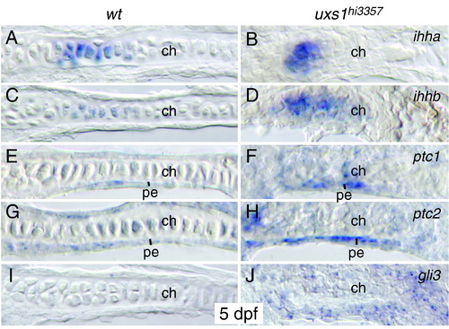
Markers of Hedgehog signaling during uxs1 mutant endochondral ossification. (A–J) In situ hybridization on horizontal sections through 5 dpf ceratohyal for ihha (A, B), ihhb (C, D), ptc1 (E, F), ptc2 (G, H), and gli3 (I, J). Wild-type expression of ihha (A) and ihhb (B) appeared similar to that observed in uxs1hi3357 mutant chondrocytes (B, D). Compared to wild-type perichondrium (E, G, I), uxs1 mutant perichondrium demonstrated increased expression of ptc1 (F), ptc2 (H), and gli3 (J). EXPRESSION / LABELING:
|
Reprinted from Developmental Biology, 341(2), Eames, B.F., Singer, A., Smith, G.A., Wood, Z.A., Yan, Y.L., He, X., Polizzi, S.J., Catchen, J.M., Rodriguez-Mari, A., Linbo, T., Raible, D.W., and Postlethwait, J.H., UDP xylose synthase 1 is required for morphogenesis and histogenesis of the craniofacial skeleton, 400-415, Copyright (2010) with permission from Elsevier. Full text @ Dev. Biol.