
- Title
-
Two tyrosine hydroxylase genes in vertebrates: New dopaminergic territories revealed in the zebrafish brain
- Authors
- Yamamoto, K., Ruuskanen, J.O., Wullimann, M.F., and Vernier, P.
- Source
- Full text @ Mol. Cell Neurosci.
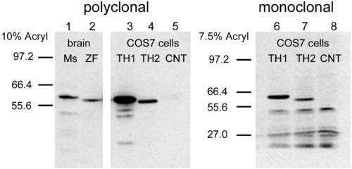
The specificity of polyclonal (left) and monoclonal (right) anti-TH antibodies for TH1 and TH2 proteins was tested on Western blots. Lanes 1 and 2 correspond to proteins extracted from mouse (Ms) and zebrafish (ZF) brains. Lanes 3 and 6 correspond to zebrafish TH1, and lanes 4 and 7 correspond to TH2, both expressed in COS7 cells. The band of the TH2 shows a lower molecular weight than TH1, and it is labeled much weaker than TH1 by the two antibodies. Lanes 5 and 8 correspond to negative controls (CNT), with lane 5 from untransfected COS7 cells, and lane 8 from COS7 cells transfected with a plasmid in which TH2 is inserted in a reverse orientation. The blot has been overexposed, in order to see the TH2 band better. |
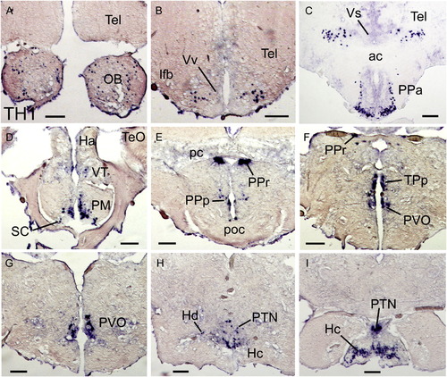
A series of transverse sections showing TH1 single ISH. TH1 transcripts were found in the olfactory bulb (A), telencephalon (Tel; B, C), preoptic area (PPa, PM, PPp; C–E), ventral thalamus (VT; D), suprachiasmatic nucleus (SC; D), pretectum (PPr; E), posterior tuberculum (TPp, PVO; F, G), posterior tuberal nucleus (PTN; H, I), and hypothalamus (Hd, Hc; H–I). See the list for abbreviations. Scale bars: 100 μm. EXPRESSION / LABELING:
|
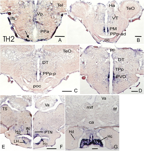
A series of transverse sections showing TH2 single ISH. TH2 transcripts were abundant in the preoptic area (PPa, PM, PPp-ad, PPp-p; A–C), the paraventricular organ (PVO; D) of the posterior tuberculum, and in the hypothalamus (LH, IN, Hd, Hc; E–G). See the list for abbreviations. Scale bars: 100 μm. EXPRESSION / LABELING:
|
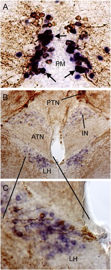
Transverse sections of preoptic (A) and hypothalamic (B and C) regions showing double-labeling for TH2 ISH (purple) and TH IHC (light brown). Double-labeled cells are shown in dark brown/purple (arrows in A), probably representing co-expression of TH1 and TH2. The hypothalamic regions (B) express predominantly TH2, with few double-labeled cells. Higher magnification of LH cells is shown in C. EXPRESSION / LABELING:
|
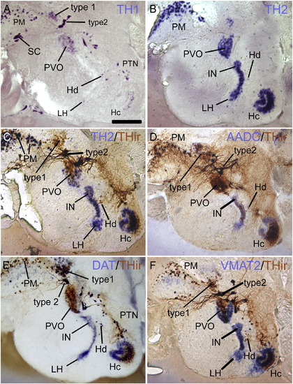
Sagittal sections through preoptic and adjacent diencephalic regions, showing TH1 single ISH (A), TH2 single ISH (B), and double-labeling for TH2 ISH (C), AADC ISH (D), DAT ISH (E), or VMAT2 ISH (F) with THir, respectively. The localization of TH1 transcript is practically identical to THir. Note that there are many double-labeled (dark brown) cells in the preoptic area (PM is shown mostly), while many TH2, AADC, DAT, and VMAT2 cells without THir are found in the hypothalamus. Scale bar = 200 μm. EXPRESSION / LABELING:
|
Reprinted from Molecular and cellular neurosciences, 43(4), Yamamoto, K., Ruuskanen, J.O., Wullimann, M.F., and Vernier, P., Two tyrosine hydroxylase genes in vertebrates: New dopaminergic territories revealed in the zebrafish brain, 394-402, Copyright (2010) with permission from Elsevier. Full text @ Mol. Cell Neurosci.