
- Title
-
Development and Regeneration of the Zebrafish Maxillary Barbel: A Novel Study System for Vertebrate Tissue Growth and Repair
- Authors
- LeClair, E.E., and Topczewski, J.
- Source
- Full text @ PLoS One
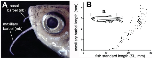
Position and growth of the paired barbels in zebrafish. A) Location of the nasal and maxillary barbels (nb and mb) on a wild type adult zebrafish (AB strain). B) Growth curve for the maxillary barbel in a wild type AB strain reared at 28°C. Barbel length (n = 183) was measured in 135 zebrafish of different standard lengths (SL+/-0.5 mm). Each data point represents a single barbel (the right and/or left appendage). The growth curve is similar to that shown in [60]. |
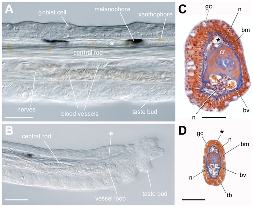
Whole-mount and sectional views of the adult maxillary barbel. A) Differential interference contrast (DIC) image of an adult maxillary barbel shaft fixed and cleared in 50% glycerol. All of the central tissues are visible through the transparent outer epithelium. A putative lymph vessel (*) lies dorsal to the central rod. All scale bars = 100 μm. B) DIC image of an adult maxillary barbel tip at the same scale as A. The central rod is reduced to a narrow band of fibers. The ventral vasculature terminates in a capillary loop packed with erythrocytes, while the lymphatic (*) terminates in a blind, tapered end. The ventral epithelium and distal tip carry numerous taste buds. C) Representative cross-section of an adult maxillary barbel at the level shown in A. Nuclei are dark red, erythrocytes orange, and basal laminae/connective tissues blue (Mallory′s trichrome). D) Representative cross-section at the level shown in B. bm = basement membrane; bv = blood vessel; gc = goblet cell; n = nerve fibers; tb = taste bud; * = putative lymph vessel (for explanation see text). |
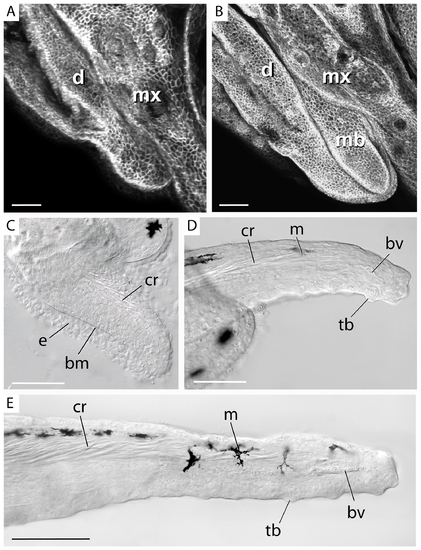
Early development of the maxillary barbel bud. A) Whole-mount confocal microscopy of the lower jaw of a juvenile membrane–GFP (mGFP) transgenic zebrafish (<10 mm standard length). Anterior is to the left. The maxillary barbel bud is not yet visible between the adjacent maxilla (mx) and dentary (d). All scale bars = 50 μm. B) Corresponding view of a slightly larger mGFP juvenile (10–12 mm SL). The first sign of the maxillary barbel (mb) is a rounded bud projecting caudoventrally. C) The early barbel bud has a thick outer epithelium (e) and a dense mesodermal core. The two layers are separated by a prominent basement membrane (bm). Fine strands of birefringent matrix accumulate dorsally where the central rod (cr) will form. D) As the barbel grows, the central rod becomes denser and projects farther distally. Isolated melanophores (m) appear along the dorsal epithelium. Ventrally, the blood vessel loop (bv) is forming. The ventral epithelium and distal tip carry numerous taste buds (tb). E) Later stages of barbel development involve expansion of the earlier structures. The central rod becomes longer and denser, the capillary loop extends, multiple melanophores become spaced along the dorsal surface, and numerous taste buds are added. The lymph vessel is not yet patent. bm = basement membrane; bv = blood vessel; cr = central rod; d = dentary; e = epithelium; m = melanophore; mb = maxillary barbel bud; ms = mesenchyme; mx = maxilla; o = orbit; tb = taste bud. |
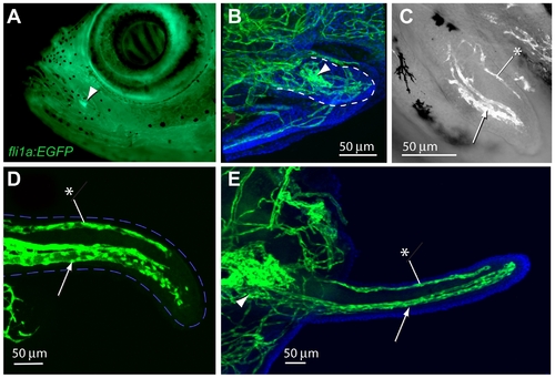
Development of the maxillary barbel vasculature in Tg(fli1a:EGFP) transgenic zebrafish. A) In vivo image of a juvenile Tg(fli1a:EGFP) zebrafish (10–12 mm standard length), in which all endothelial cells fluoresce green. The base of the future maxillary barbel is visible externally as a bright green confluence of blood vessels on the posterior ventral corner of the maxilla (arrowhead). B) 75 µm barbel. Confocal reconstruction of the early barbel bud circulation; anterior is to the left. A confluence of green vessels is visible at the base of the barbel (arrowhead). Smaller endothelial sprouts invade the bud proper (within the dotted line). Nuclei are counterstained blue (DAPI). C) 125 μm barbel. Two streams of endothelial cells are visible; a larger ventral stream (arrow), which will form the capillary loop, and a smaller dorsal stream (asterisk), which will form the putative lymphatic. In this focal plane, the proximal plexus of vessels is not visible. D) 300 μm barbel. The proximal ends of the ventral and dorsal vessels appear patent and lined with flattened endothelial cells. The distal ends of the vessels are composed of loose amoeboid cells with filipodia projecting into the surrounding tissue. The outline of the barbel is dashed blue. E) 600 μm barbel. The circulation at this stage consists of a closed capillary loop ventrally and a single, blind-end vessel dorsally. The proximal vascular plexus is greatly enlarged. Nuclei are counterstained blue (DAPI). Arrowhead = proximal vascular plexus; arrow = ventral vessels; asterisk = dorsal vessel (putative lymphatic). |
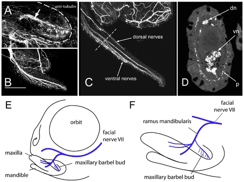
Innervation of the maxillary barbel. A) 75 μm barbel. In all panels, anterior is to the left. Whole-mount immunohistochemistry (anti-acetylated tubulin) shows a central tract of nerve fibers (arrow) within the early barbel bud (dotted white line). Smaller nerve projections are concentrated in the ventral half of the appendage. B) 200 μm barbel. Multiple fascicles of nerve fibers project distally, innervating the barbel′s ventral side and distal tip. No large tracts are visible dorsally. Scale bar = 100 μm. C) 1 mm barbel. Secondary nerve fibers appear within the dorsal half of the barbel. D) Section of an adult barbel at the approximate level shown by the dotted line in C. Innervation is visible as two deep nerve tracts (dn and vn) and a ring of sub-epithelial immunoreactive punctae (p). E,F) Schematic reconstructions of maxillary barbel bud innervation based on confocal tracing of whole-mount acetylated tubulin immunostaining in multiple zebrafish juveniles. F is an enlargement of the jaw region in E. |
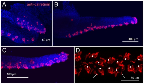
Maxillary barbel taste bud development. A) 150 μm barbel. Whole-mount immunohistochemistry (anti-calretinin) shows numerous differentiated taste buds (red) on the ventral surface and distal tip of the early barbel bud. Nuclei are counterstained blue (DAPI stain). B) 400 μm barbel. Teardrop-shaped clusters of calretinin positive cells line the ventral surface. C) Magnification of the maxillary barbel tip. D) Ventral view of the mature maxillary barbel. Teardrop-shaped taste buds (white dots) are arranged in pairs along the ventral surface. Scattered solitary chemosensory cells (SCCs, white arrows) are visible between the taste bud clusters. |
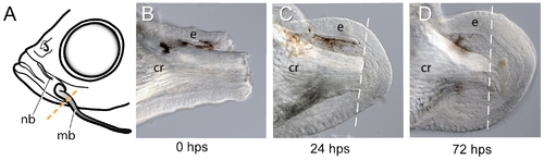
Proximal amputation of the maxillary barbel induces wound healing and blastema formation. A) Procedure for a unilateral “barbectomy”. The left maxillary barbel (mb) is amputated at the posterior margin of the maxilla. The contralateral barbel (not shown) is left as a non-surgical control. B–D) Fixed, unstained barbel regenerates collected immediately post surgery (B, 0 hps), or after 24 and 72 hours, respectively (C, D). Wound healing is followed by an accumulation of small, rounded mesenchymal cells underneath the epithelium (e) and around the central rod (cr). |
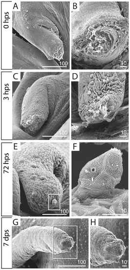
Scanning electron microscopy of early barbel regenerates. A, B) Immediately after amputation (zero hours post surgery, or 0 hps), the barbel stump is an open wound exposing the central core. A) Barbel stump in lateral view; proximal is to the left. B) End-on view of the same specimen. C, D) Two separate specimens collected at three hours post surgery (3 dps). The adjacent epithelium has closed the wound completely. Note the “purse string” lines of contraction within the epithelial sheet (C). Erythrocytes, dying cells, and matrix debris adhere to the distal surface (D). E,F) After 72 hours, the barbel stump swells distally, becoming bulbous (E). The tip epithelium carries newly differentiating taste bud hillocks, complete with protruding apical villi (F, magnified from E). G,H) By 7 days post surgery (7 dps), the maxillary barbel is a smaller version of the original appendage. Several millimeters long, it has a tapered distal end that carries a dense cluster of taste buds (H, magnified from G). |
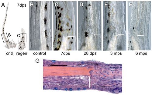
Internal scarring of maxillary barbel regenerates. A) Gross morphology of matched maxillary barbels (cntl = control; regen = regenerate) collected 7 days post surgery (dps). Note that the regenerate is thicker than the contralateral control and contains a central rod “stump.” B) Magnification of the control barbel in A. C) Magnification of the regenerated barbel in A. Note the absence of the central rod and the presence of wavy strands of matrix distal to the original amputation plane (arrow). The epithelial surface, pigment cell patterning and capillary loop are largely normal. D–F) Three regenerated barbels collected at 1–6 months post surgery (mps). All show disorganized mesodermal cores distal to the plane of section (arrow). G) Longitudinal histological section of a maxillary barbel regenerate (10 dps) showing disorganization of mesodermal cells and extracellular matrix distal to the amputation plane (arrow). Proximal is to the left. Hematoxylin/eosin stain. |
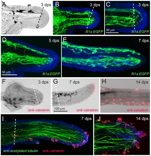
Regeneration of the barbel vasculature, taste buds, and sensory nerves. A) Transmitted light image of a Tg(fli1a:EGFP) maxillary barbel stump 3 days post surgery (dps). The distal end projects right. B) Confocal image of the vasculature in A. Endothelial sprouts (green) project distally past the plane of section and appear to bridge the dorsal (top) and ventral (bottom) vessels. Nuclei are counterstained blue (DAPI). C) Confocal reconstruction of a second regenerating barbel (3 dps). Similar sprouting is visible, as well as several isolated endothelial cells migrating underneath the wound epithelium. D–E) Regeneration of the vasculature at 5 and 7 days post surgery. Two streams of endothelial cells are visible; a dorsal stream (top) and a ventral stream (bottom). Both sets of vessels are more torturous than those of the original barbel (e.g., Fig. 4E). F–H) Regeneration of the taste buds 3–14 days post surgery (dps). Calretinin-positive cells appear at the tip within 3 days (F). By 7 dps, the distribution of taste buds on the ventral side and distal tip resembles the normal adult pattern (G). The ventral taste buds of a 14-day regenerate (H) are arranged in a typical double row (compare to Fig. 6D). I, J) Regenerating maxillary barbels are densely innervated with long axons (anti-acetylated tubulin, green) projecting to the bases of the taste buds (anti-calretinin, red). |
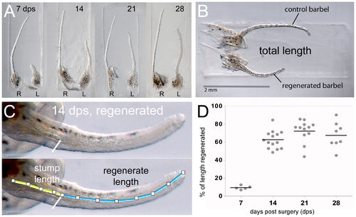
Barbel regeneration does not restore appendage length. A) Four matched pairs of maxillary barbels collected 7–28 days post surgery and embedded in the wells of a DNA electrophoresis gel. R = right barbel (unoperated control); L = left barbel (regenerate). All regenerates are shorter than the contralateral appendages. Each panel is shown at the same magnification. B) Measurement of total length (TL). The total length of each barbel (segmented line) was measured from the base of the central rod to the distal tip of the appendage. C) Measurement of stump length (SL) and regenerate length (RL). Within each regenerating barbel, the stump was measured from the base of the central rod to the amputation plane. The regenerate length was measured from the amputation plane to the distal tip. Stump length + regenerate length = total length of the regenerating barbel (SL + RL = TL). D) The regrowth of each regenerate was calculated as a percent of the control (% of length regenerated = (regenerate length/(control length – stump length))*100). Most lengthening occurred 7–14 days post surgery. Longer periods of regeneration (21–28 days) did not yield statistically significant differences in length. |
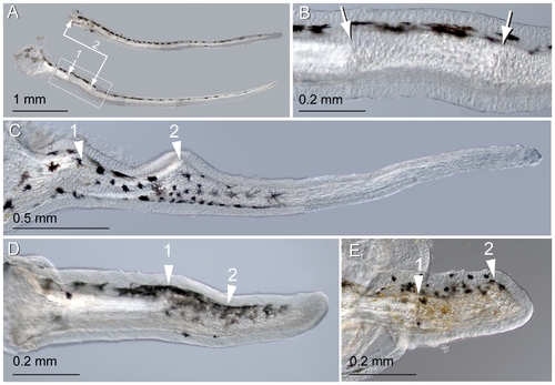
Repeated amputation can induce secondary regeneration. A) Two maxillary barbels regenerated from the same stump. The original barbel (not shown) was amputated at site 1 and the stump allowed to regenerate for one month. The resulting appendage (primary regenerate, top) was then amputated again slightly distal to the first amputation plane (site 2). A secondary regenerate (bottom) grew that was similar in size, shape and pigmentation to the primary regenerate. B) A magnification of the two surgical sites in A. Primary and secondary scars are visible approximately 0.5 mm apart. Note that the epithelial surface and melanophore patterning are largely normal. C) A secondary regenerate with more extreme scarring and swelling at the primary (1) and secondary (2) surgical sites. D–E) Failure of secondary regeneration. Secondary regenerates often failed to grow, elongating either slightly (D) or not at all (E) past the secondary surgical site (2). |
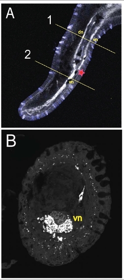
Abnormal regrowth of maxillary barbel nerve tracts A) Whole-mount immunohistochemistry of a regenerated barbel (7 dps). Nerves = white (acetylated tubulin). The red star indicates the amputation plane. Proximal to this level (1), there are two major nerves tracts, dorsal and ventral. Distal to this level (2), there is only one. dn = dorsal nerves; vn = ventral nerves. B) Section of the specimen at level 2, showing all nerve tracts displaced ventrally (vn). |