
- Title
-
Differential expression of neuroligin genes in the nervous system of zebrafish
- Authors
- Davey, C., Tallafuss, A., and Washbourne, P.
- Source
- Full text @ Dev. Dyn.
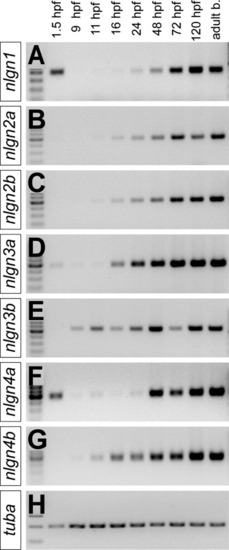
Analysis of nlgn expression levels during development. Expression levels were assayed by RT-PCR using primers specific to (A-G) the zebrafish nlgns across multiple developmental stages and in the adult brain, and (H) tubulin alpha control. From left to right, cDNA samples were derived from wild-type (AB/Tübingen) zebrafish at the 16-cell (1.5 hpf), 90% epiboly (9 hpf), 3-somite (11 hpf), 16-, 24-, 48-, 72-, and 7-dpf stages and from adult brain. |
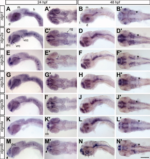
Expression of nlgns in the developing nervous system. The expression patterns for the nlgn genes were revealed by whole-mount ISH and are displayed as lateral views and dorsal views (A-N′) at 24 (A,C,E,G,I,K,M) and 48 hpf (B,D,F,H,J,L,N). d, diencephalon; e, eye; h, hindbrain; m, midbrain; t, telencephalon; cg, cranial ganglia; ob, olfactory bulb; dorso-rostral cluster; vcc, ventro-caudal cluster; vrc, ventro-rostral cluster; asterisks (*) denote the otic vesicles. Scale bar = 110 μm in 24-hpf embryos and 90 μm in 48-hpf embryos. |
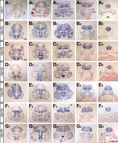
Expression of nlgns in the developing brain at 48 hpf. The expression patterns for the nlgn genes (A-G) were examined by ISH in cross-sections of embryos at 48 hpf. The levels of the sections (1-5) correspond to sections 2, 3, 5, 8, and 12, respectively, in the ZFIN atlas of zebrafish anatomy (zfin. org/zf_info/anatomy/48hrs/48hrs.html). d, diencephalon; e, eye; m, midbrain; h, hindbrain; ht, hypothalamus; hy, hypophysis; op, olfactory placode; ot, optic tectum; ov, otic vesicle; pfb, pectoral fin bud. Scale bar = 105 μm in A1-G4 and 100 μm in A5-G5. EXPRESSION / LABELING:
|
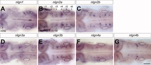
Expression of nlgns in the developing hindbrain. The expression patterns for the nlgn genes were examined by whole-mount ISH in the hindbrain of zebrafish embryos at 24 hpf. This revealed cell-specific expression patterns in large neurons of the reticulospinal tract. Individual cells located in r4, possibly Mauthner cells, are highlighted with arrows in A and G. Rhombomeres of the hindbrain are labeled with arrows in B. m, midline; r, rhombomere; cg, cranial ganglia; mhb, midbrain-hindbrain boundary. Scale bar = 50 μm. EXPRESSION / LABELING:
|
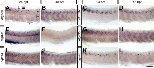
Expression of nlgns in the developing trunk. ISH performed on 24- and 48-hpf whole-mount zebrafish embryos reveals that the nlgn genes are expressed in the developing spinal cord in a dynamic and cell-specific manner. Dorsal cells that are presumably Rohon-Beard neurons are highlighted with arrows in A. s, somite; nc; notochord; sc, spinal cord. Scale bar = 35 μm. |
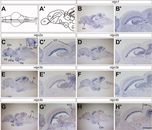
Expression of nlgns in adult brain. A: ISH staining is displayed in saggital sections of adult brain at a medial (m) and a more lateral level (l) as indicated in the dorsal view of adult brain. Medial sections are displayed in B through H and more lateral sections are displayed in B′ through H′. A′: Schematic of the forebrain displays subregions of the telencephalon. B-H: The entire brain. B′-H′: An enlargement of optic tectum, valvula, and corpus cerebelli. The inset in C is an enlargement of the thalamus and periventricular pretectal nucleus. CCe, corpus cerebelli, Cm, corpus mamillare; Dm, medial portion of the dorsal telencephalon; Hy, hypothalamus; OT, optic tectum; PP, periventricular pretectal nucleus; Th, thalamus; Tl, torus longitudinalis; Vd, dorsal portion of the ventral telencephalon; Vv, ventral portion of the ventral telencephalon; GCL, granule cell layer; PCL, Purkinje cell layer; VCe, valvula cerebelli. Scale bar = 750 μm in medial sections and 320 μm in lateral sections. EXPRESSION / LABELING:
|