
- Title
-
Molecular pedomorphism underlies craniofacial skeletal evolution in Antarctic notothenioid fishes
- Authors
- Albertson, R.C., Yan, Y.L., Titus, T.A., Pisano, E., Vacchi, M., Yelick, P.C., Detrich, H.W. 3rd, and Postlethwait, J.H.
- Source
- Full text @ BMC Evol. Biol.
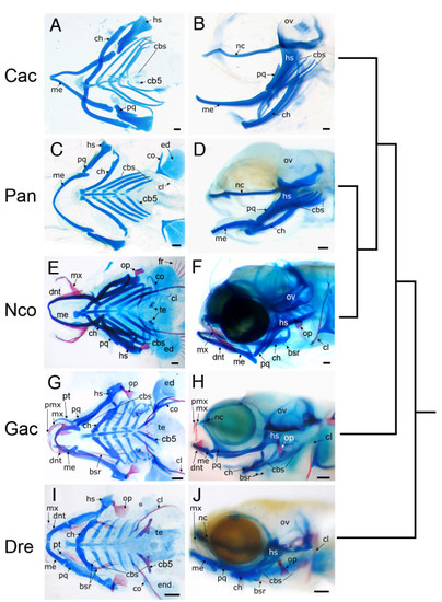
Fish Specimens. Cleared and stained skeletal preparations of the Antarctic silverfish (P. antarcticum), the blackfin icefish (C. aceratus), the yellowbelly rockcod (N. coriiceps), the threespine stickleback (G. aculeatus), and the zebrafish (D. rerio) at comparable stages of development. Bone is stained red with Alizarin red and cartilage is stained blue with Alcian blue. (A, B) C. aceratus, mid-larvae; (C, D)P. antarcticum, mid-larvae; (E, F) N. coriiceps, mid-larvae; (G, H) G. aculeatus, 10 dpf; (I, J) D. rerio, 6 dpf. Both ventral flat mounted (A, C, E, G, I) and lateral (B, D, F, H, J) views are shown. Note the lack of bony elements in pelagic notothenioid species compared to the benthic notothenioid and outgroups. The tree to the right of the images indicates evolutionary relationships. Abbreviations: bsr, branchiostegal rays; cbs, ceratobranchal cartilages; cb5, 5th ceratobrachial cartilage; ch, ceratohyal; cl, cleithrum; co, corocoid; dnt, dentary; ed, endochondral disk; fr, fin rays; hs, hyosymplectic; me, Meckel′s cartilage; mx, maxilla; nc, neurocranium; op, opercle; ov, otic vesicle; pmx, premaxilla; pq, palatoquadrate; pt, pterygoid process of the palatoquadrate; te, teeth. Scale bars, 100 μm. |
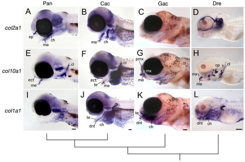
Whole-mount in situ hybridization for collagen gene expression in the pharyngeal skeleton. Lateral views are shown of embryos for P. antarcticum, C. aceratus, G. aculeatus, and D. rerio at comparable stages of development. (A, E, I) P. antarcticum, mid-larvae; (B, F, J) C. aceratus, mid-larvae; (C, G, K)G. aculeatus, 11 dpf; and (D, H, L) D. rerio, 5 dpf. Notothenioids retain strong col2a1 expression in the pharyngeal skeleton (A, B) well into larval development compared to both G. aculeatus (C) and D. rerio (D). Conversely, col10a1 (E, F) and col1a1 (I, J) expression is relatively weak or absent in the notothenioids. The tree represents the evolutionary relationship among species. Abbreviations: ch, ceratohyal; cl, cleithrum; dnt, dentary; ep, ethmoid plate; ect, ectopterygoid; me, Meckel′s cartilage; mx, maxilla; op, opercle; pmx, premaxilla; te, teeth. Scale bars, 100 μm. EXPRESSION / LABELING:
|
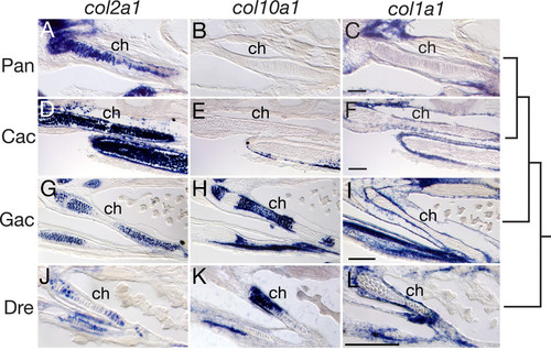
Ceratohyal collagen gene expression in P. antarcticum, C. aceratus, G. aculeatus, and D. rerio detected by in situ hybridization on cryosections. (A-C) P. antarcticum, mid-larvae; (D-F) C. aceratus, mid-larvae; (G-I) G. aculeatus, 11 dpf; (J-L) D. rerio, 6 dpf. Sections through the ceratohyal cartilage (ch) are shown in the ventral view. Notothenioid larvae show ubiquitous expression of col2a1 throughout the ch (A, D), whereas col2a1 expression is restricted to the distal ends of the cartilage in both G. aculeatus (G) and D. rerio (J). The col10a1 gene is expressed in hypertrophic chondrocytes in the ch in G. aculeatus (H) and D. rerio (K), whereas its expression is absent from the ch in P. antarcticum (B) and C. aceratus (E). The col1a1 gene is weakly expressed in the periosteum around the ch of both P. antarcticum (C) and C. aceratus (F) relative to the strong expression observed in G. aculeatus (I)and D. rerio (L). The tree represents the evolutionary relationship among species. Scale bars, 100 μm. EXPRESSION / LABELING:
|
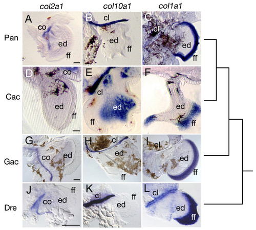
Expression of collagen genes in the pectoral fins of P. antarcticum, C. aceratus, G. aculeatus, and D. rerio embryos, detected by whole-mount in situ hybridization. (A-C) P. antarcticum, mid-larvae; (D-F) C. aceratus, mid-larvae; (G-I) G. aculeatus, 7 dpf; (J-L)D. rerio, 3 dpf. Images are of flat-mounted (A-C, G-L) or vibratome-sectioned (D-F) pectoral fins. Expression of col2a1 is observed in the scapulocoracoid (co) of all species (A, D, G, J). Likewise expression of col10a1 (B, E, H, K) and col1a1 (C, F, I, L) is observed in the cleithrum (cl), and col1a1 is expressed in the fin fold (ff) of all species. C. aceratus exhibits a unique pattern of col10a1 expression in the endochondral disk (ed) (E). Thus, gene expression during fin development appears to be largely conserved among these species. The tree represents the evolutionary relationship among species. Scale bars, 100 μm. EXPRESSION / LABELING:
|
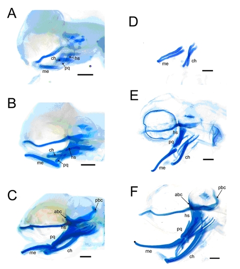
Developmental stages of P. antarcticum (A-C) and C. aceratus (D-F). (A, C) Prehatching stages, equivalent to stages ∼60 hpf and ∼52 hpf, respectively, of the zebrafish hatching period (19). (B, E) Early-larvae stages, equivalent to ∼74-90 hpf of the zebrafish larval period. Since C. aceratus specimens were systematically collected from a single clutch over the course of ∼40 day, staging was a bit more straightforward. The C. aceratus specimen in (E) is 8 days older than that depicted in (D); it also corresponds to approximately zebrafish stage ∼74 hpf. This is the protruding mouth stage in zebrafish and is characterized by the anterior extension of the jaw just past the eye. Based on these two time-points (i.e., D and E), we inferred that each day of development in notothenioids (at -1.5° C) corresponds to 2.5 hrs of development in zebrafish (at 28.5° C). (C, F) Mid-larvae stages, equivalent to zebrafish stage 5-6 dpf. The specimen in (F) is 24 days older that (E), and should therefore be at a similar stage as a 6 dpf zebrafish larvae. This is consistent with the presence of teeth in C. aceratus larvae at this stage. Staging of P. antarcticum was less straight forward, as samples represented natural populations of embryos collected beneath the sea ice (63). The sample in (A) was collected from Terra Nova Bay, Antarctica, on November 7, 2005, and was determined to be equivalent to zebrafish stage ∼60hpf. The sample in (B) was collected 20 days later, but there was clearly a range of developmental stages present in this collection. In particular, there were differences in the degree of mouth protrusion. This particular fish represented one of the younger samples, and based on the presence and absence of individual skeletal structures (Table S1), was determined to be at approximately the same stage as C. aceratusin panel (E). Fish collected on November 27 were subsequently reared for another 16 days in aquaria, and, based on the developmental “clock” applied to C. aceratus, these fish should be equivalent to zebrafish stage ∼5 dpf. As before, this collection contained a range of stages, and the specimen depicted in panel (C) was one of the older individuals equivalent to zebrafish stage ∼5.5 dpf. |