
- Title
-
Developmental and tissue-specific expression of NITRs
- Authors
- Yoder, J.A., Turner, P.M., Wright, P.D., Wittamer, V., Bertrand, J.Y., Traver, D., and Litman, G.W.
- Source
- Full text @ Immunogenetics
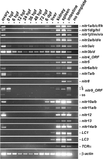
RT-PCR analyses of NITR gene expression from zebrafish ovaries, embryos, and adult tissues. Zebrafish (AB strain) embryos were collected by natural mating and maintained at 28°C as described (Westerfield 2000). Total RNA (2 μg) from pooled embryos at various developmental stages (hours post-fertilization (hpf) or days post fertilization (dpf)) and dissected tissues (pooled from multiple animals) was reverse transcribed (SuperScript™ II Reverse Transcriptase: Invitrogen; Carlsbad, CA) and subjected to thermal cycling with gene-specific primers (Table 1 and Fig. 1) and TITANIUM™ Taq DNA polymerase (Clontech). Primers that amplify the entire open reading frame were utilized for nitr4 and nitr9: note that three mRNA splice variants of nitr9, (Wei et al. 2007) are detected in the kidney and intestine. RT-PCR of immunoglobulin light chains (LC1 and LC3), T cell receptor α (TCR-α) and β-actin are included for reference. TCR-α primers were designed to amplify sequences between the variable (V) and constant (C) domains and thus only detect transcripts, which reflect somatic recombination. The degenerate TCR-α forward primers were designed to detect all V domain sequences encoding the conserved motif: A(V/L)YYCA. Limited RNA blot analyses with polyA+ mRNA from intestine, kidney and spleen, detected transcripts between 1.4-2.0 Kb for the zebrafish nitr1-nitr7 families (unpublished observations) |
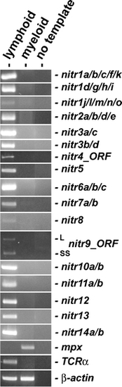
RT-PCR analyses of NITR gene expression from different leukocyte populations. Lymphoid and myeloid cell populations were purified from the kidney of multiple zebrafish and pooled as described (Yoder et al. 2007). RT-PCR was performed as described in Fig. 2, except 1 μg of total RNA was used in the reverse transcription reaction. RT-PCR of myeloperoxidase (mpx) provides a positive control for myeloid cells and TCR-α provides a positive control for T lymphocytes. β-actin is shown as a standard reference. Primer sequences and thermal cycling parameters are listed in Table 1 EXPRESSION / LABELING:
|