
- Title
-
Canonical Wnt signaling is required for the maintenance of dorsal retinal identity
- Authors
- Veien, E.S., Rosenthal, J.S., Kruse-Bend, R.C., Chien, C.B., and Dorsky, R.I.
- Source
- Full text @ Development
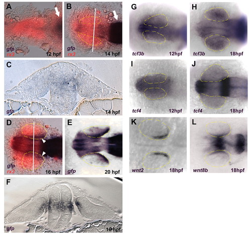
Wnt signaling becomes active in the dorso-posterior retinal pigmented epithelium (RPE) between 14 and 16 hpf. (A-F) Expression of the TOP:dGFP Wnt reporter detected using in situ hybridization for gfp (blue). In A, B and D, the embryos were also probed for rx3 expression (red) which marks the eye field. (A,B) Dorsal views, anterior left. Active Wnt signaling does not extend rostrally past the midbrain-hindbrain boundary (arrows) at 12 hpf, and approaches but does not enter the eye field at 14 hpf. (C,F) Coronal sections through caudal midbrain/posterior optic vesicles, dorsal up. The lines in B and D indicate the planes of section in C and F, respectively. Active Wnt signaling is seen in the dorsoposterior RPE at 16 hpf, but not at 14 hpf. (D,E) Dorsal views, anterior left. Active Wnt signaling is clearly present in the dorso-posterior eye field at 16 and 20 hpf. (G-L) Dorsal views, anterior left. (G-J) Expression of tcf3b and tcf4 is present in the early eye-field during optic vesicle evagination (12 hpf) and throughout the eye at 18 hpf. (K) The Wnt ligand wnt2 is expressed in the dorsal RPE at 18 hpf. (L) Expression of wnt8b in the midbrain and RPE at 18 hpf. EXPRESSION / LABELING:
|
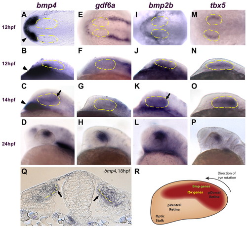
Multiple Bmp genes and tbx5 are expressed in the retina before canonical Wnt activity. (A,E,I,M) Dorsal views, anterior left. (B-D,F-H,J-L,N-P) Lateral views, dorsal up, anterior left. (A-D) bmp4 is expressed in the prechordal mesoderm at 12 and 14 hpf (arrowheads in A-C) but is not expressed in the optic vesicle until 14 hpf (arrow in C). At 24 hpf, bmp4 expression is restricted to the dorsal retina (D). (E-L) gdf6a and bmp2b are not expressed in the optic vesicle at 12 hpf (expression of these genes is restricted to the surface ectoderm). Expression of bmp2b is present in the retina at 14 hpf (arrow in K), but gdf6a does not appear in the optic vesicle until 16 hpf (not shown). Both genes are expressed in the dorsal retina at 24 hpf (H,L). (M-P) tbx5 expression begins in the optic vesicle at 12 hpf and becomes progressively restricted to the dorsal retina by 24 hpf. (Q) Transverse section through the midbrain at 18 hpf. bmp4 is expressed in the presumptive dorsal neural retina and RPE (arrows). Broken yellow lines indicate the interface between the neural retina and the RPE. (R) Diagram of the zebrafish retina at approximately 14 hpf, showing the expression domains of Bmp genes and tbx5 at this timepoint. At approximately 22 hpf, the entire eye rotates 90° in the direction indicated. Anterior left, dorsal up. EXPRESSION / LABELING:
|
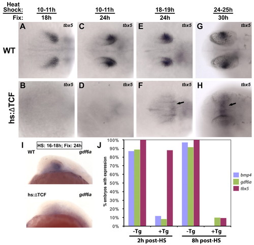
Expression of dorsal retinal genes is lost following the repression of Wnt signaling. The Tg(hsp70l:Tcf3-GFP)w26 transgenic zebrafish line, which expresses a dominant-repressor form of Tcf3 (ΔTcf-GFP) upon heat shock, was used for these experiments. Dorsal views, anterior left. (A-H) Embryos were heat shocked and fixed at the indicated times, and sorted for GFP expression. The repression of Wnt targets led to the downregulation of tbx5 in the dorsal retina at every timepoint. tbx5 expression was upregulated in the dorsal diencephalon at later timepoints (arrows in F,H). (I) Expression of gdf6a was also eliminated in embryos expressing ΔTcf at 18 hpf and fixed at 24 hpf. (J) To determine the times at which bmp4, gdf6a and tbx5 are lost in the dorsal retina following the repression of Wnt targets, embryos were heat shocked at 16 hpf and fixed 2 and 8 hours later. bmp4 and gdf6a were strongly reduced at the 2 hour timepoint, whereas tbx5 was still expressed. By 8 hours, the expression of all three genes was lost. EXPRESSION / LABELING:
|
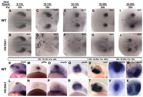
Wnt signaling is required for the maintenance of dorsal retinal identity. The Tg(hsp70l:dkk1-GFP)w32 transgenic zebrafish line, which expresses the secreted Wnt pathway inhibitor Dkk1 upon heat shock, was used for these experiments. (A-J) Dorsal views, anterior left. Embryos were heat shocked and fixed at the indicated times, and sorted for GFP expression. (A,B) Embryos fixed just before Wnt signaling becomes active in the dorsal RPE express tbx5 normally, showing that tbx5 expression initiates properly in the absence of Wnt signaling. (C-J) Inhibition of Wnt signaling caused a strong downregulation of tbx5 at the early timepoints, with a weaker effect at the last timepoint. This demonstrates a requirement for Wnt signaling in the maintenance of tbx5. (K-T) Lateral views, dorsal up, anterior left. (K-P) Expression of the Bmp ligands bmp4, gdf6a and bmp2b are lost from the dorsal retina following Wnt inhibition, suggesting a loss of dorsal character. (Q-T) pax6b is expressed normally and vax2 expands dorsally, suggesting a ventralized retina. (U-X) Whole eyes, dorsal up. Following Wnt inhibition, the expression of ephrin B2a (efnb2a) is downregulated in the dorsal retina, but maintained in the lens, and ephb2 expands dorsally. EXPRESSION / LABELING:
|
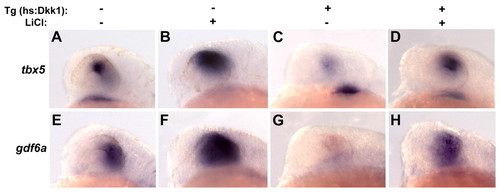
Activation of Wnt signaling rescues loss of dorsal eye markers in Dkk1-expressing embryos. (A-H) Dkk1-expressing embryos were treated with the Wnt pathway activator LiCl (150 mM) from 11-14 hpf, heat shocked at 12 hpf, and fixed at 24 hpf. The expression of tbx5 (A-D) and gdf6a (E-H) were analyzed by in situ hybridization. LiCl led to an expansion of tbx5 and gdf6a expression in embryos not expressing Dkk1 (B,F), and a rescue of tbx5 and gdf6a in embryos expressing Dkk1 (D,H). Lateral views, dorsal up, anterior left. EXPRESSION / LABELING:
|
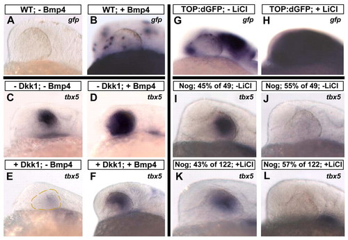
Bmp signaling is downstream of Wnt signaling in the maintenance of dorsal retinal markers. (A-F) Bmp4 can rescue dorsal retinal markers in the absence of Wnt signaling. hs:Dkk1 and control wild-type (WT) embryos at the one-cell stage were injected with a construct that expresses Bmp4 upon heat shock (pDestTol2pA2;hsp70l:bmp4-IRES-GFP), heat shocked at 12 hpf, and fixed at 24 hpf. (A,B) To illustrate transgene expression following heat shock, in situ hybridization was performed for gfp. Widespread clonal expression was observed in the retinas in 85% of embryos. (C-F) Expression of Bmp4 caused a clear expansion of tbx5 in embryos not expressing Dkk1 (D) and the rescue of tbx5 in embryos expressing Dkk1 (F). (G-L) The activation of Wnt signaling does not rescue dorsal markers in the absence of Bmp signaling. Embryos heterozygous for the Tg(hsp70l:nog3)fr14 transgene, which express the Bmp pathway inhibitor Noggin upon heat shock, were outcrossed to TL strain fish and placed in 200 mM LiCl at 18 hpf. A 2-hour heat shock was performed at 18 hpf, and embryos were fixed at 24 hpf. To illustrate Wnt pathway activation, TOP:dGFP embryos were similarly treated with LiCl from 18-24 hpf and gfp detected by in situ hybridization (G,H). For hs:Noggin embryos untreated with LiCl, 55% of 49 embryos lost expression of tbx5 (J) and, for embryos treated with LiCl, 57% of 122 embryos lost expression of tbx5 (L), showing that the activation of Wnt signaling cannot rescue tbx5 in the absence of Bmp signaling. Lateral views, dorsal up, anterior left. EXPRESSION / LABELING:
|