
- Title
-
The interaction of nucleoside diphosphate kinase B with Gbetagamma dimers controls heterotrimeric G protein function
- Authors
- Hippe, H.J., Wolf, N.M., Abu-Taha, I., Mehringer, R., Just, S., Lutz, S., Niroomand, F., Postel, E.H., Katus, H.A., Rottbauer, W., and Wieland, T.
- Source
- Full text @ Proc. Natl. Acad. Sci. USA
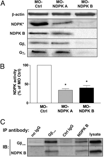
NDPK A and B depletion in zebrafish embryos. Zebrafish embryos were injected with MO-Ctrl (4 ng), MO-NDPK A (8 ng) and MO-NDPK B (4 ng). (A) Immunoblot analysis of lysates prepared 72 hpf using specific antibodies against total fish NDPK (NDPK*), NDPK B, Gβ1 and Gγ2. β-actin served as loading and specificity control. (B) Relative NDPK activity (normalized to MO-Ctrl) was quantified as formation of 3H-GTP from 3H-GDP and ATP in lysates of embryos injected as indicated. Data are means ± SEM, n = 3, *, P < 0.05 vs. MO-Ctrl. (C) Co-immunoprecipitation of NDPK B and Gβ in zebrabrafish lysates. Endogenous Gβ or NDPK B was immunoprecipitated (IP) from zebrafish lysates. Association of NDPK B or Gβ was detected by immunoblotting (IB). Representative immunoblots are shown. EXPRESSION / LABELING:
PHENOTYPE:
|
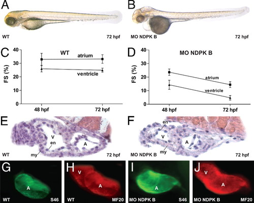
Cardiac dysfunction in NDPK B-depleted zebrafish embryos. Cardiac phenotype of wild-type (WT) (A) and NDPK B knockdown (B) embryos at 72 hpf. Fractional shortening (FS) of the atrial and ventricular chamber of WT (C) and NDPK B morphants (D) at 48 and 72 hpf. Data are means ± SEM, n = 11–21. Histology sections of a WT (E) and NDPK B knockdown (F) heart region stained with hematoxylin/eosin. A, atrium, V, ventricle, en, endocardial, my, myocardial layer. (G–J) Whole-mount immunofluorescence following staining with specific antibodies against atrial myosin heavy chain [S46, (G) and (I)] and entire heart tube myosin heavy chain [MF20, (H and J)] showing unaltered expression of structure proteins. EXPRESSION / LABELING:
PHENOTYPE:
|
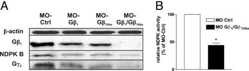
Gβ1 and Gβ1like depletion in zebrafish embryos. Zebrafish embryos were injected with MO-Ctrl (4 ng), MO-Gβ1 (3 ng) and MO-Gβ1like (3 ng) singly or in combination (3 + 3 ng). (A) Immunoblot analysis of lysates prepared 72 hpf using specific antibodies against NDPK B, Gβ1 and Gγ2. β-actin served as loading and specificity control. (B) Relative NDPK activity (normalized to MO-Ctrl) was quantified as formation of 3H-GTP from 3H-GDP and ATP in lysates of embryos injected as indicated. Data are means ± SEM, n = 3, *, P < 0.05 vs. MO-Ctrl. EXPRESSION / LABELING:
PHENOTYPE:
|
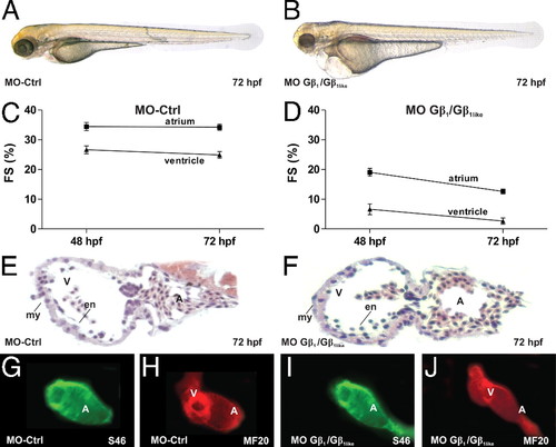
Loss of zebrafish Gβ1 phenocopies NDPK B morphants. Cardiac phenotype of control morpholino-injected (MO-Ctrl) (A) and MO-Gβ1/Gβ1like double-injected (B) embryos at 72 hpf. Fractional shortening (FS) of the atrial and ventricular chamber of MO-Ctrl fishes (C) and Gβ1/Gβ1like morphants (D) at 48 and 72 hpf. Data are means ± SEM, n = 10–12. Histology sections of a MO-Ctrl (E) and Gβ1/Gβ1like knockdown (F) heart region stained with hematoxylin/eosin. A, atrium, V, ventricle, en, endocardial, my, myocardial layer. (G–J) Whole-mount immunofluorescence following staining with specific antibodies against atrial myosin heavy chain [S46, (G and I)] and entire heart tube myosin heavy chain [MF20, (H and J)] showing unaltered expression of structure proteins. EXPRESSION / LABELING:
PHENOTYPE:
|
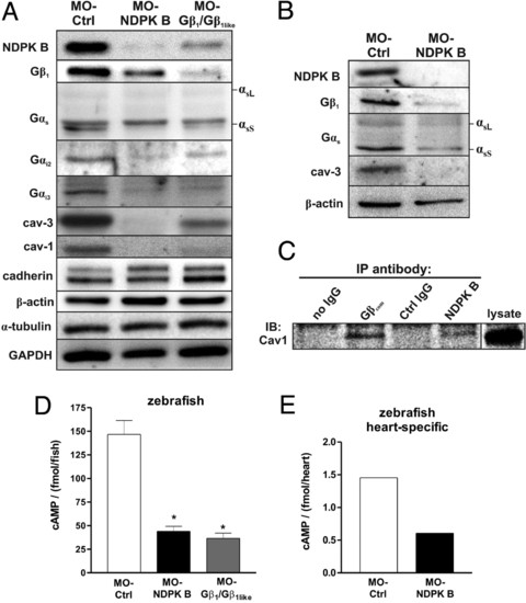
Alterations of G protein content and cAMP formation in NDPK B and Gβ knockdown zebrafish embryos. (A and B) Representative immunoblots of zebrafish embryos at 72 hpf following knockdown of either NDPK B or Gβ1/Gβ1like in total fish lysates (A) and heart-specific extracts (B) using specific antibodies against the indicated G protein subunit and caveolin isoform. Positions of the long (αsL) and short (αsS) Gαs splice variants are indicated. Note that GαsS is migrating below a nonspecific immunoreactive band. Marker proteins (cadherin, β-actin, α-tubulin, GAPDH) representing different cell compartments are shown as loading and specificity control. (C) Co-immunoprecipitation of caveolin-1 with NDPK B and Gη. Endogenous Gβ or NDPK B was immunoprecipitated (IP) from zebrafish lysates (refer to Fig. 1C). Association of caveolin-1 was detected by immunoblotting (IB). A representative immunoblot is shown. (D) cAMP was measured in lysates from whole embryos at 72 hpf injected with the indicated MOs. Data are means ± SEM, n = 3, *, P < 0.05 vs. MO-Ctrl. (E) cAMP content of zebrafish hearts isolated from MO-Ctrl vs. MO-NDPK B-injected fishes with heart-specific GFP expression [Tg(cmlc2:gfp)] at 72 hpf. n = 365 hearts were pooled and lysed for each condition, respectively. The average of two independent experiments is shown. EXPRESSION / LABELING:
PHENOTYPE:
|
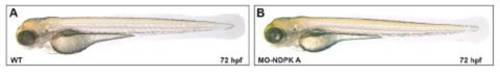
NDPK A knockdown in zebrafish embryos. (A and B) Phenotype (lateral view) of wild-type (WT) (A) and NDPK A knockdown (B) embryos at 72 hpf after injection of 8 ng MO-NDPK A. |
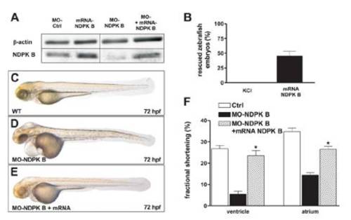
Rescue in NDPK B morphants. Zebrafish embryos were injected with MO-Ctrl (4 ng), NDPK B-mRNA (0.8 ng) and MO-NDPK B (4 ng) as well as co-injected with both MO-NDPK B (4 ng) and NDPK B-mRNA (0.8 ng) that lacks theMObinding site. (A) Representative immunoblots of zebrafish lysates prepared at 72 hpf confirmed mRNA driven NDPK B expression. (B) Quantitative analysis of rescued NDPK B morphants (4 ng) by co-injection of 0.8 ng NDPK B-mRNA according to phenotypic appearance. Data are means±SEM, n=3, 70–80 fishes respectively. Representive phenotypes ofWT(C),NDPKB morphants (D) and rescued embryos (E). (F) Rescue of cardiac contractility. Fractional shortening of the ventricular and atrial chamber of WT, NDPK B morphants and rescued morphants are shown. Data are means ± SEM, n = 10–15 for each condition, *, P < 0.05 vs. MO-NDPK B. |
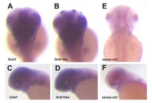
Pattern of Gβ1 and Gβ1like mRNA content in zebrafish embryos as analyzed by in situ-hybridization. Expression pattern of Gβ1 (A, dorsal view; C, lateral view) and Gβ1like (B, dorsal view; D, lateral view) mRNA in zebrafish embryos at 72 hpf as detected by whole mount in situ hybridization. Dorsal (E) and lateral view (F) of sense RNA probed embryos as a control. To avoid cross reactions despite high homology, digoxygenin-incorporated antisense RNA probes were directed against sequences in the 5′-untranslated region of the zebrafish gnb1 and gnb1l gene, respectively, in which both genes are largely different. Whole-mount in situ hybridization revealed an identical pattern of Gβ1 or Gβ1like mRNA content. |
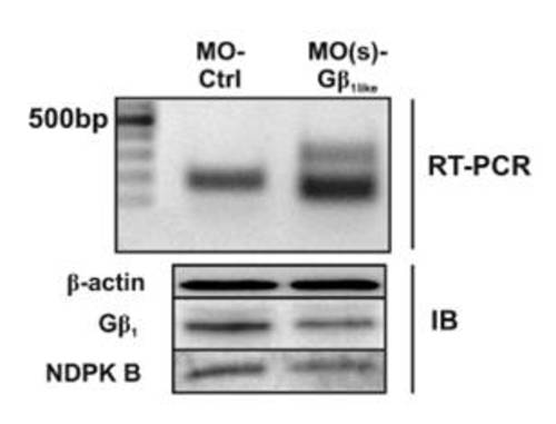
Splice morpholino-mediated knockdown of Gβ1like. Zebrafish embryos were injected with MO-Ctrl or the splice morpholino MO(s)-Gβ1like. Transcripts of Gβ1like mRNA were amplified by RT-PCR (Upper) and the protein content of NDPK B, Gβ1, and β-actin was analyzed by immunoblot (Lower). |