
- Title
-
A role for nephrin, a renal protein, in vertebrate skeletal muscle cell fusion
- Authors
- Sohn, R.L., Huang, P., Kawahara, G., Mitchell, M., Guyon, J., Kalluri, R., Kunkel, L.M., and Gussoni, E.
- Source
- Full text @ Proc. Natl. Acad. Sci. USA
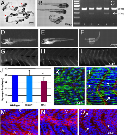
Nephrin morpholino experiments in zebrafish reveal smaller muscles. (A) Light micrograph of variable phenotypes noted with nephrin morpholino in zebrafish embryos at 4 days post fertilization (dpf). Many of the embryos appear curved and shorter (red arrows) compared with control (black arrow). (B) Light micrograph of wild-type (top) mismatched (middle) and nephrin morpholino embryos (MO1) at 4 dpf. (C) RT-PCR analysis of mismatched and nephrin morpholino-injected embryos confirms the expected splicing defect leading to an in-frame deletion of the transmembrane domain, rendering a protein no longer able to anchor at the membrane; this is manifest by a decrease in RT-PCR product size (from 398 bp to 272 bp). Lane 1, molecular weight markers; lane 2, uninjected wild-type embryo; lane 3, 1.25 ng nephrin morpholino MO1; lane 4, 1.25 ng mismatched morpholino; lane 5, 2.5 ng nephrin morpholino MO1; lane 6, 2.5 ng mismatched morpholino. (D-I) Morphometric analyses performed on whole-mount embryos stained with anti β-dystroglycan antibody confirm that the myosepta to myosepta distance of embryos injected with nephrin morpholino MO1 (F, I) are smaller than those injected with mismatched morpholino (E, H) or wild-type uninjected embryo (D, G). (J) Bar graph of myosepta length measurements from wild-type, mismatched MO1 and nephrin morpholino MO1 injected embryos. Asterisk denotes a statistically significant P value by t test (P d 0.00000004). Individual measurements are listed in Table S1. Whole-mount control (K) and nephrin morpholino (L) embryos stained with anti-laminin (red) and anti-myosin heavy chain antibody (green); nuclei are stained in blue with DAPI. Nephrin morpholinos displayed shorter myosepta and presence of clustered nuclei (white arrows). (M-O) Whole-mount 1 dpf embryos are stained with anti-β-catenin antibody (red), and nuclei are stained in blue with DAPI. (M) Wild-type control; (N) control mismatched morpholino (MISMO1); (O) nephrin morpholino (MO1). Open arrows in (M) and (N) point to nuclei that appear regularly aligned, whereas arrows in (O) point to clustered nuclei. |