
- Title
-
Identification of zebrafish A2 adenosine receptors and expression in developing embryos
- Authors
- Boehmler, W., Petko, J., Woll, M., Frey, C., Thisse, B., Thisse, C., Canfield, V.A., and Levenson, R.
- Source
- Full text @ Gene Expr. Patterns
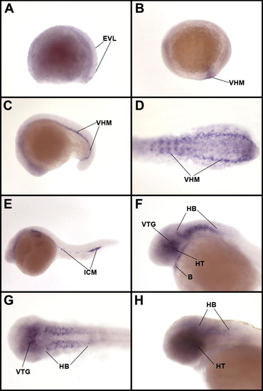
Expression of the adora2a.1 gene. Lateral view of embryos at (A) gastrula stage, (B) early somitogenesis (11 hpf), (C) mid-somitogenesis (18 hpf), (D) dorsal view of a flat mounted embryo at 18 hpf in the caudal region. Lateral view of embryos at (E) 24 hpf, (F) 36 hpf, (G) dorsal view of an embryo at 36 hpf after dissecting off the yolk. (H) lateral view of embryo at 48 hpf. B, blood; EVL, enveloping layer; HB, hindbrain; HT, hypothalamus; ICM, inner cell mass; VHM, ventral hematopoietic mesoderm; VTG, ventral tegmentum. EXPRESSION / LABELING:
|
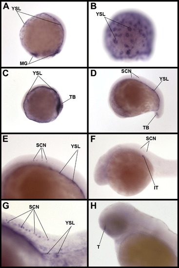
Expression of the adora2a.2 gene. Lateral view of embryos at (A) gastrulation, (B) gastrulation, focussing on the lateral part of the YSL and showing accumulation of transcripts in perinuclear area of the YSL, (C) early somitogenesis (11 hpf), (D–E) mid-somitogenesis (15 hpf) with (E) focussing on the trunk region of the embryo, (F–G) 24 hpf, with (G)focussing on the trunk region, (H) 48 hpf. IT, interrenal tissue; MG, margin; SCN, spinal cord neurons; T, telencephalon; TB, tail bud; YSL, yolk syncytial layer. EXPRESSION / LABELING:
|
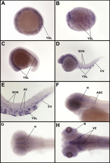
Expression of the adora2b gene. (A) Lateral view of embryo at early somitogenesis (11 hpf), (B) Dorsal view of embryo at early somitogenesis (11.5 hpf) showing labeling in YSL nuclei. Lateral view of embryos at (C) mid-somitogenesis (15 hpf), (D) 24 hpf, (E) 24 hpf in the trunk region, (F) 48 hpf. Dorsal view of the head region at (G) 48 hpf, (H) 5 dpf. ASC, anterior spinal cord; AV, axial vasculature; CV, caudal vein; H, hindbrain; SCN, spinal cord neurons; R, retina; T, telencephalon; VZ, ventricular zone; YSL, yolk syncytial layer. EXPRESSION / LABELING:
|

Caffeine protects dopaminergic neurons from MPTP-induced neurotoxicity. Dorsal views of 5 dpf embryos hybridized with dat probe. Embryos were treated with drug from 24 hpf through 5 dpf. n = 30 embryos/treatment group. (A) Untreated control embryo. (B) Embryo treated with 40 μM MPTP. (C) Embryo treated with 10 μM caffeine and 40 μM MPTP. (D) Embryo treated with 10 μM caffeine. Neurons of the ventral diencephalon are circled. Black arrows point to the bilateral pretectal cluster of neurons. EXPRESSION / LABELING:
|

Unillustrated author statements |
Reprinted from Gene expression patterns : GEP, 9(3), Boehmler, W., Petko, J., Woll, M., Frey, C., Thisse, B., Thisse, C., Canfield, V.A., and Levenson, R., Identification of zebrafish A2 adenosine receptors and expression in developing embryos, 144-151, Copyright (2009) with permission from Elsevier. Full text @ Gene Expr. Patterns