
- Title
-
Requirement of vasculogenesis and blood circulation in late stages of liver growth in zebrafish
- Authors
- Korzh, S., Pan, X., Garcia-Lecea, M., Winata, C.L., Pan, X., Wohland, T., Korzh, V., and Gong, Z.
- Source
- Full text @ BMC Dev. Biol.
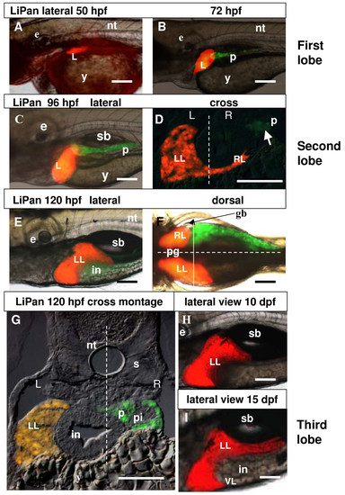
Liver development in LiPan transgenic zebrafish. (A, B) Initial RFP expression in the liver starts at 48–53 hpf (A) and GFP expression in the exocrine pancreas starts at 67–72 hpf (B). (C) Expression of RFP and GFP at 96 hpf. (D) Cross section of a 96 hpf larvae shows RFP-positive liver. GFP-expressing exocrine pancreas is faintly visible (arrow). The dotted line represents the midline of the larva and left (L) and right (R) sides are indicated. (E, F) Expression of RFP and GFP in the larva at 120 hpf: lateral (E) and dorsal view (F). The dotted horizontal line in (F) represents the midline with the right side at the top. The solid vertical line represents the plan of the cross section in (G). (G) A cross section to illustrate morphology of internal organs and expression of transgenes right (R) sides are indicated. (H, I) Lateral view of RFP-expressing liver at 10 dpf (H) and in a 120-hpf larva. The dotted line represents the midline of the larva and left (L) and 15dpf (I). Abbreviations: e, eye; gb, gall bladder; in, intestine; L, liver; LL, left lobe; RL, right lobe; nt, notochord; p, pancreas; pi, principal islet; pg, pigment; s, somite, VL, ventral lobe. In all whole mount images anterior is towards the left. Scale bars, 125 μm. EXPRESSION / LABELING:
|
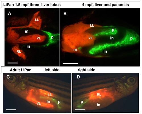
Liver and pancreas in adult LiPan fish. (A, B) Ventral view of the three liver lobes (red) in the dissected LiPan zebrafish at 1.5 mpf (A) and 4 mpf (B). The three lobes of liver are indicated by LL, left lobe; RL, right lobe; and VL, ventral lobe. The exocrine pancreas (p) is in green. (C, D) lateral view of LiPan adult: the left side (C) and the right side (D). Other abbreviation: in, intestine. Scale bars, 1500 μm. EXPRESSION / LABELING:
|
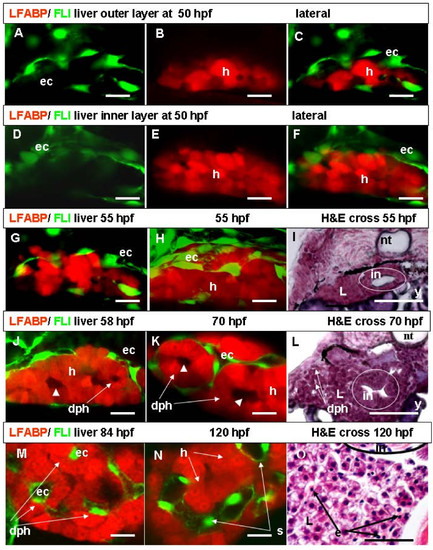
Liver vasculogenesis. (A-C) Left-lateral confocal live images of liver of LiPan/Tg(fli1:EGFP)y1 larva at 50 hpf. Discrete ECs frame outer layers of the liver bud (A-C) while no ECs are present in the inner layer (D-F). Panels A and D are images under a GFP filter, panels B and E under a RFP filter, and panels C and F are combinations of GFP and RFP images. (G, H) Left-lateral confocal live images of LiPan/Tg(fli1:EGFP)y1 liver at 55 hpf: external liver layers (G) and all liver layers (H). ECs establish contact and enter superficial hepatocytes layer. (I) Hematoxylin and eosin (H&E) staining of a cross section of an embryo at 55 hpf to show tightly packed hepatocytes inside the liver. (J, K, M, N) Confocal sections of internal liver layers at 58 hpf (J), 70 hpf (K), 84 hpf (M) and 120 hpf (N). At 58 hpf, RFP negative areas in the innermost liver layers (arrowhead) and a daisy pattern of hepatocytes are formed around this areas (J). At 70 hpf, the daisy pattern of hepatocytes around RFP negative areas extended to the superficial layers and ECs surround the daisy clusters of hepatocytes from outside (K). In some cases, ECs are inside of daisy clusters of hepatocytes (M). By 120 hpf, the size of sinusoids significantly increases (N). (L, O) H&E staining of cross sections of embryos at 70 hpf (L) and 120 hpf (O). Note a daisy pattern of hepatocytes in (L) and erythrocytes with pink cytoplasm in liver sinusoids (O). Abbreviations: dph, daisy pattern of hepatocytes; ec, endothelial cells; e, erythrocyte; h, hepatocyte; in, intestine; nt, notochord; L, liver; s, sinusoid; y, yolk. In all whole mount images anterior is towards the left. Scale bars represent 625 μm except for Panels (I, L, O), where the scale bars are 125 μm. |
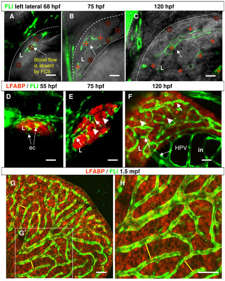
Assessment of blood circulation in the developing liver of zebrafish. (A-C) Left lateral 3D confocal sections of liver at the level of sinusoids where blood flow was measured. In the most outer liver layers where the first sinusoids were formed, blood flow was absent at 68 hpf (A). Blood flow was detected in sinusoids of external dorso-lateral part of liver parenchyma at 75 hpf (B) and in all measured sites of liver sinusoids at 120 hpf (C). Red cross, points of measurement at the different focal distance as deep as 70–80 μm from the liver surface; "No" sign, points in sinusoids where no blood flow was detected. The edges of livers are marked by dash lines. Arrows indicate the focused sinusoids for FCS measurement in the picture. (D-F) Confocal projections show three stages of liver vasculogenesis. ECs start to contact the surface layer of hepatocytes in the liver bud at 55 hpf (D), first sinusoids form between external layers of hepatocytes at 75 hpf (E) and have well developed sinusoidal network at 120 hpf (F). Arrows are endothelial cells and sinusoids; arrowheads are clusters of hepatocytes. (G, H) Confocal images demonstrate the sinusoidal network (green) in the liver of 1.5-month-old fish. (H) is a 2x blow-up of the area defined by the white box (G′) to show the sinusoidal network and two rows of hepatocytes between two neighboring sinusoids as indicated by yellow lines. Abbreviations: ec, endothelial cells; in, intestine, L, liver; HPV, hepatic portal vein. In all images anterior is towards the left-hand side. Scale bars are 625 μm in (A-B) and 300 μm in (D-H). EXPRESSION / LABELING:
|
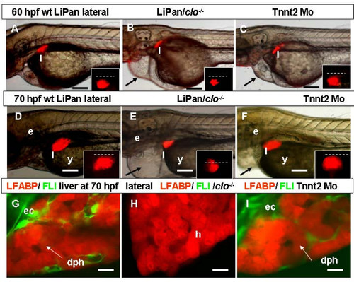
Role of endothelia in liver development. (A-F) Left-lateral views: liver morphogenesis in live LiPan/Tg(fli1:EGFP)y1 larvae; wild type (A, D), LiPan/clo-/- mutants (B, E) and LiPan/Tnnt2 morphants (C, F) at 60 hpf (A-C) and 70 hpf (D-F). Dorsal views of the liver region of the same embryos are shown as inserts in each panel and dash lines indicate the midline with the right side at the top. Note that the liver in LiPan/clo-/- mutants is significantly reduced compared to that in controls and Tnnt2 morphants. The pericardial edemas in both clo-/- mutant and tnnt2 morphant are indicated by arrows (B, C, E, F). (G-I) Left-lateral confocal in vivo projections of liver of LiPan/Tg(fli1:EGFP)y1 larvae in wild type (G), clo-/- mutant (H) and Tnnt2 morphant (I) backgrounds. Abbreviations: bd, bile duct; dph, daisy pattern of hepatocytes; ec, endothelial cells; e, ear; h, hepatocytes; l, liver; s, sinusoid. In all images, anterior is towards the left. Scale bars, 125 μm in (A-F) and 625 μm in (G-I). |
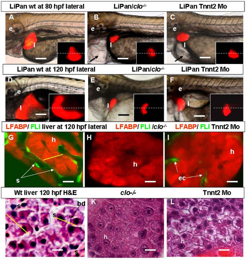
Role of circulation in liver development. (A-F) Left-lateral views: liver morphogenesis in live LiPan wild type (A, D), LiPan/clo-/- mutants (B, E) and LiPan/Tnnt2 morphants (C, F) at 80 hpf (A-C) and 120 hpf (D-F). Dorsal views of the liver region of the same embryos are shown as inserts in each panel and dash lines indicate the midline with the right side at the top. Livers in clo-/- mutants and Tnnt2 morphants are located more medial, lack the anterio-ventral and posterior expansion, and are significantly reduced in size compared to the livers in wild type sibling. The cardiac edema in both mutants and morphants is indicated by arrow. (G-I) Left-lateral confocal in vivo projections of liver of LiPan/Tg(fli1:EGFP)y1 larvae at 120 hpf in wild type (G), clo-/- mutant (H) and Tnnt2 morphant (I) backgrounds. In clo-/ mutants ECs are absent (H), whereas in tnnt2 morphants they are present only between hepatocytes of the outer layer (I). (J-L) High-resolution light micrographs of hepatic parenchyma of zebrafish larvae stained with H&E. In 120-hpf wild type sibling, hepatocyte tubules are separated by sinusoids containing erythrocytes (J); in contrast, in 120-hpf clo-/- mutant (K) and Tnnt2 morphant (L), hepatocytes are tightly connected to each other. Note two sinusoids separated by two rows of neighboring hepatocytes as defined by yellow lines. Abbreviations: ec, endothelial cells; e, ear; h, hepatocytes; l, liver; s, sinusoid. In all images, anterior is towards the left. Scale bars, 125 μm in (A-F) and 625 μm in (G-L). EXPRESSION / LABELING:
PHENOTYPE:
|