FIGURE SUMMARY
Title

Development and notch signaling requirements of the zebrafish choroid plexus

Authors
Bill, B.R., Balciunas, D., McCarra, J.A., Young, E.D., Xiong, T., Spahn, A.M., Garcia-Lecea, M., Korzh, V., Ekker, S.C., and Schimmenti, L.A.
Source
Full text @ PLoS One

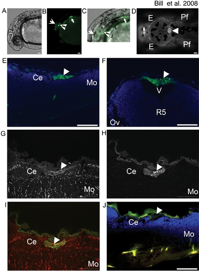
EtMn16 co-localizes with Gfap in mCP epithelia.
GFP is expressed in multiple anatomical locations in EtMn16 larvae (A–D). At 30 hpf (A–C)(lateral orientation), GFP is expressed in cells of the otic vesicle (inverted arrowhead), the pectoral fin (closed arrow), ventral hindbrain and spinal cord (open arrow) brightfield image (A), fluorescent image (B), and merged image (C). In addition to cells of the ventral hindbrain, at 4 dpf (D)(dorsal orientation), two additional structures express GFP, the diencephalic CP (dCP) (closed arrow), and the myelencephalic CP (mCP) (closed arrowhead). The mCP lies posterior to the cerebellum (Ce) [(5 dpf, 6 μm longitudinal cryosection) (E)], within the ventricle (v) dorsal to the fifth rhombomere (r5)[5 dpf, 6 μm transverse cryosection (F)]. Cells of the mCP express Gfap (G) and colocalize with GFP expressing cells (H), merged image (I) and are not seen in the negative control lacking antibodies for Gfap and GFP (J). Abbreviations: eye (E), pectoral fin (Pf), cerebellum (Ce), and medulla oblongata (Mo). All images except F are oriented anterior to the left; F is oriented dorsal to the top. Scale bar is 50 μm.

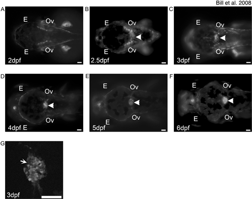
Development of the mCP as defined by EtMn16 larvae.
Fluorescent images of dorsal-oriented live zebrafish taken at 2 dpf (A), 2.5 dpf (B), 3 dpf (C), 4 dpf (D), 5 dpf (E), and 6 dpf (F); arrowhead indicates mCP. GFP-expressing cells first appear diffusely across the roof plate of the fourth ventricle (B), as defined by the level of the otic vesicles (Ov). Cells migrate toward the midline and finish coalescence by 4 dpf. The expression in this structure remains static through 6 dpf. Not all cells of the mCP express GFP (G, arrow). In all images, anterior is to the left, and scale bar is 50 μm. Abbreviations: eye (E), otic vesicle (Ov).

EXPRESSION / LABELING:
Gene:
Fish:
Anatomical Terms:
Stage Range: Pec-fin to Day 6

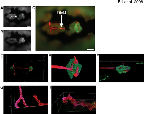
The Dorsal Longitudinal Vein supplies the mCP with its vascular network.
The Dorsal Longitudinal Vein (DLV) closely associates with the mCP epithelia as defined by doubly transgenic EtMn16/Tg(gata1:dsRed) dorsally-mounted 5 dpf live zebrafish, epithelia express GFP (A) and red blood cells express DsRed (B) merged image (C) (scale bars are 50 μm). The vessels, as defined by blood flow, directly contact the dorsal surface of the mCP. Low magnification of a 5 dpf dorsally mounted live EtMn16/Tg(gata1:dsRed), high magnification three-dimensional renders of DLV/PCeV junctions and TCB of zebrafish shown in D (E,F). Panel F is rotated 80° into the plane to show association (dorsal facing the top of the image). The DLV bends dorsally slightly before the bifurcation, traverses the mCP epithelial domain, and then turns ventrally as it transitions to the PCeV and PCeV′. Panels G and I are three-dimensional renders of the DLV, PCeV and TCB [(G,I – 85° rotation into the plane)(oriented with anterior to the right)]. The mCP has been digitally removed, and the DLV has been bisected utilizing the clipping tools of Image4D in order to define the vascular structure. Scale is as indicated on each render.

The DLV develops via angiogenic sprouting and supplies the mCP.
The development of the integral vasculature in the mCP was examined in living zebrafish larvae. The DLV (arrow in all panels) sprouts from the MCeV/MCeV′ (gray open arrowheads) via angiogenic sprouting between 40 hpf (A) and 48 hpf (B), and develops via growth cone-like filopodial extensions (C). The PCeV and PCeV′ (white open arrowheads) grow dorsomedially to meet the DLV in the roof of the fourth ventricle (D). Fusion occurs between the DLV (arrow) and PCeV or PCeV′ (E), followed by extension to the symmetric partner (PCeV or PCeV′) (F). Once connected, the DLV branches once more (G, small arrowhead) to form the trans-choroid plexus branch (TCB) (small arrowhead). By 120 hpf, the main vasculature of the mCP is in place (H). A diagrammatic representation of the final structure, with naming of the vessels and a superimposed mCP for comparison is shown (I). Small connecting vessels (concave arrow) connecting the DLV and TCB elaboration to form the mCP (J). Abbreviations: mesencephalic vein (MsV & MsV′), middle cerebral vessel (MCeV and MCeV′), dorsal longitudinal vein (DLV), posterior cerebral vein (PCeV and PCeV′) and myelencephalic choroid plexus (mCP). All images are oriented anterior to the left, and scale bars are 50 μm.

EXPRESSION / LABELING:
Gene:
Fish:
Anatomical Terms:
Stage Range: Long-pec to Day 5

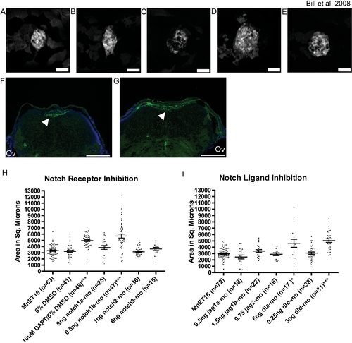
Notch signaling is required for proper development of the myelencephalic choroid plexus.
Pan Notch Inhibition with DAPT (B, dorsal-mounted live larvae and G, transverse section) results in an increase in the mCP epithelial domain compared to vehicle-treated control larvae (A, dorsal mounted live larvae and F, transverse section). This increase in domain size is due to lateral spreading as the mCP remains as a monolayer (G, DAPT-treated versus F, vehicle treated). Further analysis showed that this effect is mediated by inhibition of notch1b (D, 5 dpf live larvae) dla, and dld (E, 5 dpf live larvae). Inhibition of notch1a (C, 5 dpf live larvae) did not significantly alter the size of the mCP epithelial domain but did effect overt structure. Panels A–F are dorsal views with anterior to the left, and panels G and H are 6 μm cryosections labeled with an antibody against GFP (green) and DAPI (blue) staining the nuclei. Quantitative measurements show the distribution mCP sizes in individual fish. Measurements are shown for Notch receptor inhibition by DAPT and morpholino experiments (H), and for Notch ligand inhibition by morpholinos (I) Each point in the histograms represents a measurement of a live larval zebrafish mCP. The mean±s.e.m. is indicated by the line and error bars respectively. Significant effects on mCP size are observed for 10 μM DAPT, notch1b, dla, and dld knockdown (* p = 0.02 and *** p<0.0001). For a full list of mean, s.e.m., and p-values, see table S2. Abbreviations: eye (E), pectoral fin (Pf), and otic vesicle (Ov). Arrows indicate mCP. Scale bars are 50 μm.

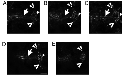
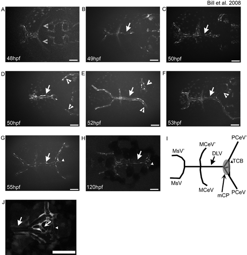
Variation of the DLV-PCeV junction. The DLV (arrows) bifurcated to join the PCeV and PCeV′ (open arrowheads) and the TCB (small arrowheads) connects the two junctions. This structure is plastic. The most common structure includes a third branch that transits between the DLV and the TCB (A–C), but in some instances the third DLV-TCB branch does not form by 5 dpf (D). The most rare occurrence is that the DLV does not develop (E). The PCeV and PCeV′ still meet in the location of the pCP, but lack blood flow (E, open arrowheads). All images are shown with anterior to the left. Scale bar is 50 μm.

EXPRESSION / LABELING:
Gene:
Fish:
Anatomical Terms:
Stage: Day 5
Acknowledgments
This image is the copyrighted work of the attributed author or publisher, and ZFIN has permission only to display this image to its users. Additional permissions should be obtained from the applicable author or publisher of the image. Full text @ PLoS One