FIGURE SUMMARY
Title

Emergence of Xin demarcates a key innovation in heart evolution

Authors
Grosskurth, S.E., Bhattacharya, D., Wang, Q., and Lin, J.J.
Source
Full text @ PLoS One

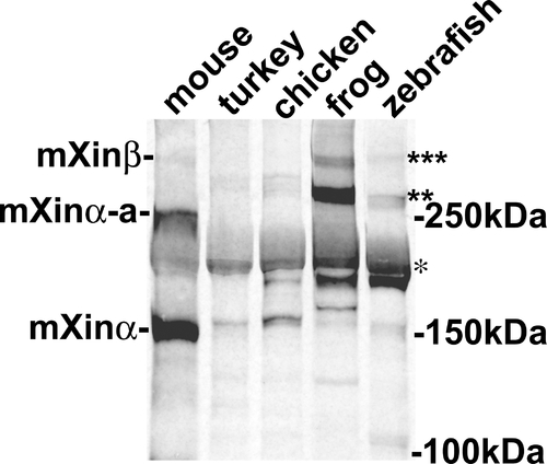
Western blot analysis of protein extracts prepared from mouse, turkey, chicken, frog and zebrafish hearts with polyclonal U1013 anti-Xin antibody.
As previously reported [5], the U1013 antibody generated against the N-terminal fragment of mXinα including the Xin repeat region specifically reacts with mXinα (155 kDa), mXinα-a (250 kDa) and mXinβ (∼340 kDa) from mouse heart. Similarly, this antibody recognizes a 217 kDa band (indicated by *), and 280–295 kDa bands (indicated by**) from turkey, chicken, frog and zebrafish heart extracts. These bands may represent Xinα and its splicing variants Xinα-a. Many degraded fragments were also detected by this antibody. In frog and zebrafish but not turkey and chicken heart extracts, this antibody also reacts with a 335 kDa band (indicated by ***), which may represent Xinβ isoform.

EXPRESSION / LABELING:
Genes:
Antibody:
Fish:
Anatomical Term:
Stage: Adult

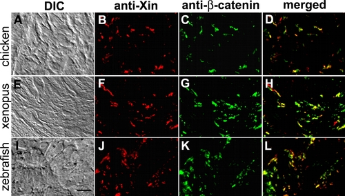
Co-localization of Xin proteins and β-catenin in chicken, frog and zebrafish hearts.
Double-label indirect immunofluorescence microscopy was performed on frozen sections of chicken (A–D), frog (E–H) and zebrafish (I–L) hearts with mouse monoclonal anti-β-catenin (C, G, K) and rabbit polyclonal U1013 anti-mXin (B, F, J) antibodies, and subsequently with a mixture of rhodamine-conjugated goat anti-rabbit IgG and fluorescein-conjugated goat anti-mouse IgG. Merged images (D, H, L) indicate co-localization of the two proteins. The differential interference-contrast (DIC) images correspond to the fluorescent images shown in respectively. Scale bar: 10 μm.

EXPRESSION / LABELING:
Gene:
Antibody:
Fish:
Anatomical Term:
Stage: Adult

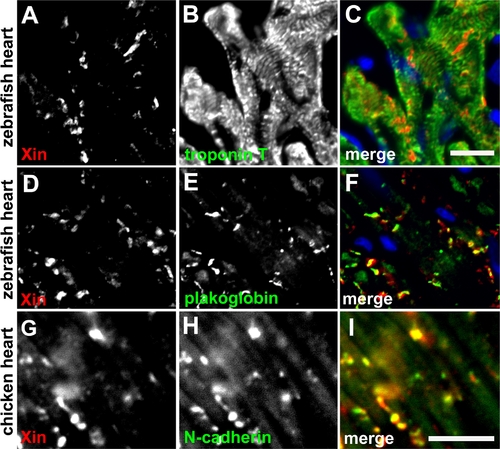
Immunofluorescence microscopy of zebrafish and chicken heart sections.
(A–F) Double-label indirect immunofluorescence was performed on frozen sections of zebrafish heart with monoclonal anti-troponin T or anti-plakoglobin (green, visualized by fluorescein-conjugated 2nd antibody) and polyclonal anti-mXin (red, visualized by rhodamine-conjugated 2nd antibody). Before mounting, the sections were treated with DAPI to stain nuclei (blue). Scale bar: 10 μm. (G–I) Double-label indirect immunofluorescence was performed on chicken heart section with monoclonal anti-N-cadherin (green) and polyclonal anti-mXin antibody (red). Scale bar: 10 μm.

Acknowledgments
This image is the copyrighted work of the attributed author or publisher, and ZFIN has permission only to display this image to its users. Additional permissions should be obtained from the applicable author or publisher of the image. Full text @ PLoS One