
- Title
-
Notch signaling regulates endocrine cell specification in the zebrafish anterior pituitary
- Authors
- Dutta, S., Dietrich, J.E., Westerfield, M., and Varga, Z.M.
- Source
- Full text @ Dev. Biol.
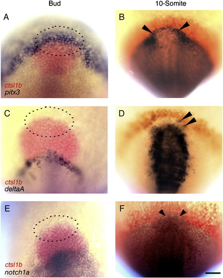
Prospective pituitary precursors express deltaA and notch1a after the 10-somite (14 h) stage. Wild-type embryos doubly labeled with mRNA probes for cathepsin L, 1b (ctsl1b), previously known as hatching gland gene 1 (hgg1; A–F, red) and pitx3 (A, B), deltaA (C, D), or notch1a (E, F). (A, C, E) At Bud stage, presumptive pituitary precursor cells express pitx3 (A, n = 25; dotted lines indicate approximate location according to Dutta et al., 2005). deltaA (C, n = 30) and notch1a (E, n = 30) are not expressed in pituitary precursors. At 10-somite stage (14 h), anterior cranial placode precursors (arrowheads) express pitx3 (B, n = 25), deltaA (D, n = 25), and notch1a (F, n = 25). (A, C, E) Dorsal views, anterior to the top (dotted lines indicate approximate location of pituitary precursor cells according to Dutta et al., 2005); (B, D, F) dorsal views of prospective head region, anterior to the top. Scale bar: 50 mμm. EXPRESSION / LABELING:
|
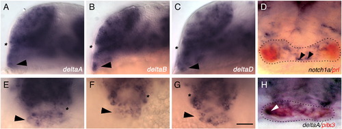
At the 20-somite (19 h) stage, pituitary placode cells express deltaA, deltaB, and deltaD in a salt and pepper pattern. Wild-type embryos are labeled with probes for deltaA (A, E, n = 19), deltaB (B, F, n = 28), and deltaD (C, G, n = 25). (D) Expression of notch1a (blue, n = 20) and prl (red, n = 20) does not colocalize in the pituitary placode at prim-5 stage (24 h). (H) Cells in the pituitary placode co-express pitx3 (red, n = 20) and deltaA (blue, n = 20). (A–H) Arrowheads indicate cells in the pituitary placode, asterisk indicates optic recess. (A–C) Side views, anterior to the left, dorsal to the top; (D–H) frontal views, dorsal to the top. Scale bar: 50 μm (A–C, E–G), 20 μm (D, H). EXPRESSION / LABELING:
|
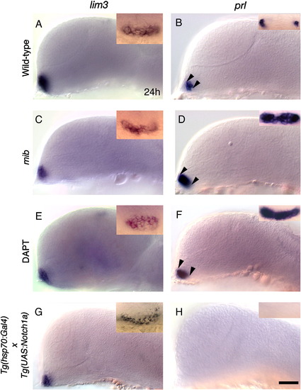
Notch is necessary and sufficient to restrict lactotrope differentiation in the anterior pituitary placode. (A, B) Wild-type embryos labeled with lim3 (A, n = 85) and prl (B, n = 96). (B, inset) Bilateral cells in the anterior pituitary placode express prl. Gene expression in DMSO treated (n = 30), and hsp70:Gal4 heat-shocked (n = 30) control embryos was similar to wild-type embryos in A and B. (A, C, E, G) Pituitary placode size and lim3 expression in mib mutant (C, n = 39), in DAPT treated (E, n = 45), and in heat-shocked hsp70:Gal4 × UAS:notch1a (G, n = 22) transgenic embryos is similar to controls (A, B). (B, D, F) Loss of functional Notch signaling in mib mutant (D, inset, n = 37/37) and DAPT treated (F, inset, n = 32/40) embryos lead to expansion of prl in pituitary placode (arrowheads; D, F). (H) prl expression in the anterior pituitary placode is completely lost in hsp70:Gal4 x UAS:notch1a transgenic embryos (n = 48/48) following heat shock. (A–G) side views, anterior to the left, dorsal to the top; (A–G, inset) frontal views, dorsal to the top. All embryos Prim-5 (24 h) stage. Scale bar: 50 μm (A–H); Insets A, C, E, G: 40 μm; Insets B, D, F, H: 30 μm. EXPRESSION / LABELING:
|
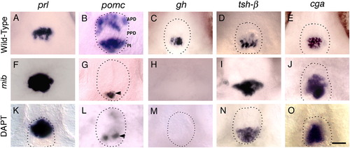
Notch signaling is necessary for corticotrope and somatotrope differentiation. (A–E) Wild-type (Gene expression in DMSO treated control embryos, not shown, was same as in wild-type embryos in panels A–E), mib mutant (F–J) and DAPT treated embryos (K–O) labeled with probes for anterior pituitary hormones at 72 h. In mib mutant embryos, prl (F, n = 25/25), tsh-β (I, n = 23/23) and cga (J, n = 25/25) in the anterior pituitary is expanded, pomc expression is lost in APD and reduced in PPD (G, n = 26/26), and gh expression (H, n = 52/52) is completely lost compared to prl (A, n = 37), tsh-β (D, n = 40) and cga (E, n = 40), pomc (B, n = 20), gh expression (C, n = 40) in siblings. DAPT treatment between shield stage and 72 h leads to expanded prl (K, n = 26/33), tsh-β (n = 50/60) and cga expression (O, n = 35/42), whereas pomc expression in APD (L, n = 23/25) is lost or severely reduced, pomc expression in PI (L, n = 22/25) is somewhat reduced and gh expression (M, n = 35/40) is entirely absent in the PPD compared to wild-type embryos (A–E). Dotted lines indicate outline of anterior pituitary tissue (omitted in panels where pituitary border was ambiguous). Arrowhead shows pomc expression in PI. (A–O) Ventral views, anterior to the top. Scale bar: 25 μm (A–O). EXPRESSION / LABELING:
|

Gain of Notch function increases the number of pomc expressing corticotropes and melanotropes at the expense of lactotropes and thyrotropes. Embryos carrying either the hsp70:Gal4 or the hsp70:Gal4 x UAS:notch1a transgenes were heat shocked at Bud (10 h) stage and analyzed for hormone expression at Protruding mouth (72 h) stage (A–J). In heat-shocked hsp70:Gal4 x UAS:notch1a transgenic embryos, both prl (F, n = 36/36) and tsh-β (I, n = 42/42) are completely lost, pomc expression is expanded in APD, PPD and PI (G, n = 32/32), and cga is expanded (J, n = 15/17). gh expression is unaffected (H, n = 40/40) compared to prl (A, n = 34) and tsh-β (D, n = 32), pomc (B, n = 37), gh (C, n = 30) and cga (E, n = 20) in hsp70:Gal4 embryos following heat shock. Dotted lines indicate outline of anterior pituitary tissue (omitted in panels where pituitary border was ambiguous). (A–J) Ventral views anterior to the top. Scale bar: 15 μm (A–J). EXPRESSION / LABELING:
|

Unillustrated author statements |
Reprinted from Developmental Biology, 319(2), Dutta, S., Dietrich, J.E., Westerfield, M., and Varga, Z.M., Notch signaling regulates endocrine cell specification in the zebrafish anterior pituitary, 248-257, Copyright (2008) with permission from Elsevier. Full text @ Dev. Biol.