
- Title
-
Expression of trpC1 and trpC6 orthologs in zebrafish
- Authors
- Möller, C.C., Mangos, S., Drummond, I.A., and Reiser, J.
- Source
- Full text @ Gene Expr. Patterns
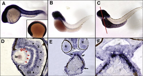
Expression of zebrafish trpC1 by whole mount in situ hybridization and histological analysis. Expression of trpC1 mRNA is ubiquitous in six somite embryos (A; inset) and stages up to and including 24 hpf (A). At 56 hpf, expression is restricted to the head with no detectable expression in the trunk. (B) Strong head expression of trpC1 persists until 72 hpf (C), in addition to expression in the outflow tract of the heart (white arrowhead; white line denotes plane of section in panels E and F). Histological examination of 72 hpf embryos reveals specific expression of trpC1 in the ganglion cell layer of the eye (gcl, red arrowheads) and in the inner nuclear layer (inl, black arrowheads). Anterior sections of 72 hpf embryos (line in C) confirm expression of trpC1 in the outflow track (E, black arrows). A magnified view (F) shows a high level of expression in the cells associated with the outflow tract (white arrowheads). Le, lens; e, eye; gcl, ganglion cell layer; inl, inner nuclear layer. EXPRESSION / LABELING:
|
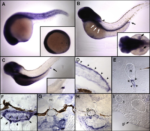
Expression of zebrafish trpC6 by whole mount in situ hybridization and histological analysis. Expression of trpC6 mRNA is ubiquitous at six somites (A, inset) and in all stages tested up to and including 24 hpf (A). (B) At 48 hpf, expression becomes restricted to the head, pectoral fins (black arrowhead), the area of the gut (white arrows) extending to the posterior end (black arrow). Dorsal view of expression in fins at 48 hpf (B, inset). This pattern of trpC6 expression persists to 72 hpf (C), where expression in the most posterior region of the gut (black arrow and inset) remains high while more proximal regions of the gut show diminished expression. Histological examination reveals that trpC6 expression in the pectoral fins is restricted to the dorsal surface (D, black arrowheads). Sectioning of the trunk of 72 hpf embryos shows that trpC6 mRNA is expressed in cells lining the dorsal aorta (E, black arrowheads). A closer examination of the gut reveals that trpC6 is highly expressed in cells that surround and encapsulate the gut (F, black arrows). (G) Sections through the glomerulus of a 3 dpf larva (G, dashed black circle) demonstrate that trpC6 RNA is not detectably expressed podocytes, whereas cells encapsulating the anterior gut are positive for trpC6 (G, white arrowheads). Later stage, 5 dpf (H) and adult glomeruli (I) do not display specific labeling for trpC6. nc, notochord; gl, glomerulus; t, tubules. EXPRESSION / LABELING:
|
Reprinted from Gene expression patterns : GEP, 8(5), Möller, C.C., Mangos, S., Drummond, I.A., and Reiser, J., Expression of trpC1 and trpC6 orthologs in zebrafish, 291-296, Copyright (2008) with permission from Elsevier. Full text @ Gene Expr. Patterns