- Title
-
Transient axonal glycoprotein-1 (TAG-1) and laminin-alpha1 regulate dynamic growth cone behaviors and initial axon direction in vivo
- Authors
- Wolman, M.A., Sittaramane, V., Essner, J.J., Yost, H.J., Chandrasekhar, A., and Halloran, M.C.
- Source
- Full text @ Neural Dev.
|
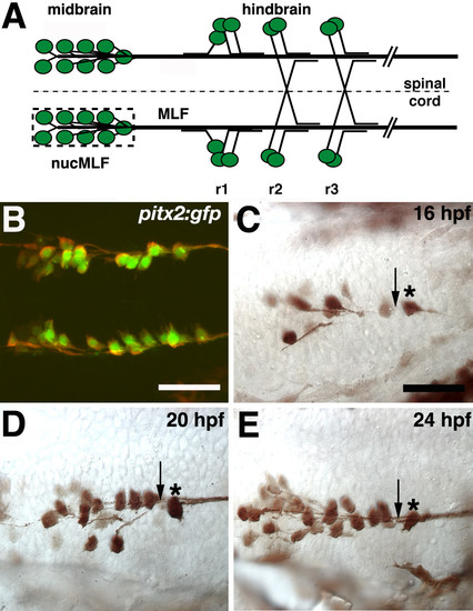
Initial extension and convergence of MLF axons.(a-e) Ventral views, anterior to the left. (a) Schematic representation of MLF axons and hindbrain axons that grow along the MLF. The dashed line denotes the ventral midline and the dashed box surrounds the 'nucMLF zone'. r = rhombomere. (b) Confocal projection of 20 hpf Tg(pitx2c:gfp) embryo stained with anti-GFP (green) and ZN-12 antibodies (red). (c-e) Whole mount preparations of Tg(pitx2c:gfp) embryos stained with anti-GFP at 16 (c), 20 (d), and 24 (e) hpf. Midline is up. Asterisks denote the caudal-most nucMLF cell and arrows indicate the 'convergence point'. Scale bar = 25 μm. EXPRESSION / LABELING:
|
|
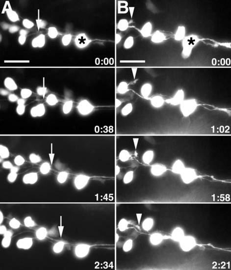
MLF growth cones fasciculate along neighboring MLF axons and are inhibited by surrounding tissue.(a, b) Images from time-lapse sequence of MLF axon outgrowth in uninjected Tg(pitx2c:gfp) embryos. Ventral views, anterior to the left, midline is up. Asterisks denote the caudal-most nucMLF cell. (a) The arrows indicate the position of the growth cone that fasciculates along a neighboring MLF axon. (b) The arrowheads label the growth cone that is repelled by surrounding tissue. The time stamp shows hours: minutes. Scale bar = 25 μm. EXPRESSION / LABELING:
|
|
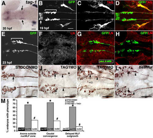
TAG-1 or laminin-α1 loss of function disrupts normal MLF axon convergence.(a-l) Ventral views, anterior to the left. (a) In situ hybridization for tag-1 in 20 hpf embryo. Arrows indicate expression in nucMLF. (b-d) Confocal projections of 19 hpf Tg(pitx2c:gfp) embryos labeled with anti-GFP (green) and anti-TAG-1 (red) antibodies. The arrow indicates unidentified cells in the diencephalon expressing the pitx2c:gfp transgene. The brackets indicate the extent of nucMLF. (e-h) Single confocal plane of nucMLF region in 23 hpf Tg(pitx2c:gfp) embryos labeled with anti-GFP (green) and anti-laminin (red). (e-g) The focal plane is at the ventral surface of the neuroepithelium. The inset in (g) is a 90 degree rotation of a z-reconstruction, with ventral down, showing the laminin underlying the nucMLF. (h) The focal plane is 4 μm dorsal to the plane in (e-g), and shows that laminin is not concentrated within the neuroepithelium. (i-l) Whole mount preparations of STDCONMO injected embryos (i), TAG1MO injectnderlying the nucMLF. (h) The focal plane is 4 μm dorsal to the plane in (e-g), and shows that laminin is not concentrated within thed embryos (j, k), and bal;Tg(pitx2c:gfp) embryos (l) labeled with an anti-GFP antibody. The outline defines the ′nucMLF zone′. Arrows indicate the normal convergence point. Filled arrowheads denote the actual convergence point. Open arrowheads label examples of MLF axons outside of the nucMLF zone. The asterisk indicates stunted MLF extension. (m) Quantification of the average percentage of embryos with MLF phenotypes. *P < 0.001, two sample binomial comparison versus STDCON. #P < 0.001, two sample binomial comparison versus STDCON. #P < 0.001, two sample binomial comparison versus TAG1MO. N equals the number of embryos. Scale bars = 50 μm. The scale bar in (b) is for (b-h). EXPRESSION / LABELING:
PHENOTYPE:
|
|
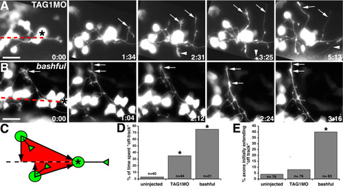
TAG-1 or laminin-α1 loss of function causes misdirected MLF axon outgrowth.(a, b) Images from timelapse sequence of MLF axon outgrowth in TAG1MO injected (a) and bal;Tg(pitx2c:gfp) (b) embryos. Ventral views, anterior to the left, midline is up. Asterisks denote the caudal-most nucMLF cell. The dashed red line marks the nucMLF centerline. Arrows indicate MLF axons that wander into surrounding tissue and do not converge within timelapse duration. Arrowheads label an MLF axon that extends into lateral tissue, but eventually converges. The time stamp shows hours: minutes. (c) Schematic representation defining on-track (red shaded region) versus off-track regions. (d, e) Quantification of the average percentage of time MLF axons grew off-track (d) and the average percentage of axons that initially emerged from their cell body in an off-track position (e). *P < 0.001, two sample binomial comparison versus uninjected. N equals the number of MLF axons. Scale bar = 25 μm. EXPRESSION / LABELING:
PHENOTYPE:
|
|
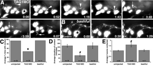
TAG-1 knockdown causes defects in MLF axon-axon interactions.(a, b) Images from time-lapse sequence of MLF axon outgrowth in TAG1MO injected (a) and bal;Tg(pitx2c:gfp) (b) embryos. Ventral views, anterior to the left, midline is up. Asterisks denote the caudal-most nucMLF cell. Arrowheads label a growth cone that repeatedly samples neighboring axons, but fails to maintain these interactions. Arrows label a growth cone that contacts another MLF axon, subsequently retracts, and then emerges from the opposite side of the neuron. The time stamp shows hours: minutes. (c-e) Quantification of the average percentage of time each growth cone contacted another MLF axon after contact was initiated (c), the average duration of these contacts (d), and the average number of axons each growth cone contacted (e). *P < 0.001, two sample binomial comparison versus uninjected. #P < 0.001, two-tailed t-test versus uninjected. Error bars represent the standard error of the mean. N equals the number of axons (c, e) and growth cone-axon contacts (d). Scale bar = 25 μm. EXPRESSION / LABELING:
PHENOTYPE:
|
|
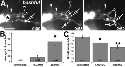
bal embryos have excessively branched axons and MLF growth rates are reduced by TAG-1 or laminin-α1 loss of function.(a) Images from timelapse sequence of MLF axon outgrowth in a bal;Tg(pitx2c:gfp) embryo. Ventral views, anterior to the left, midline is up. The asterisk denotes the caudal-most nucMLF cell. Open and filled arrows and arrowheads label branches from individual axons. The time stamp shows hours: minutes. (b, c) Quantification of the average number of branches per axon (b) and the average growth rate of MLF axons (c). #P < 0.001, Kruskal-Wallis ANOVA versus uninjected. *P < 0.05, **P < 0.001, two-tailed t-test versus uninjected. Error bars represent the standard error of the mean. N equals the number of axons. Scale bar = 25 μm. EXPRESSION / LABELING:
PHENOTYPE:
|

