
- Title
-
Zebrafish cdx1b regulates expression of downstream factors of Nodal signaling during early endoderm formation
- Authors
- Cheng, P.Y., Lin, C.C., Wu, C.S., Lu, Y.F., Lin, C.Y., Chung, C.C., Chu, C.Y, Huang, C.J., Tsai, C.Y., Korzh, S., Wu, J.L., and Hwang, S.P.
- Source
- Full text @ Development
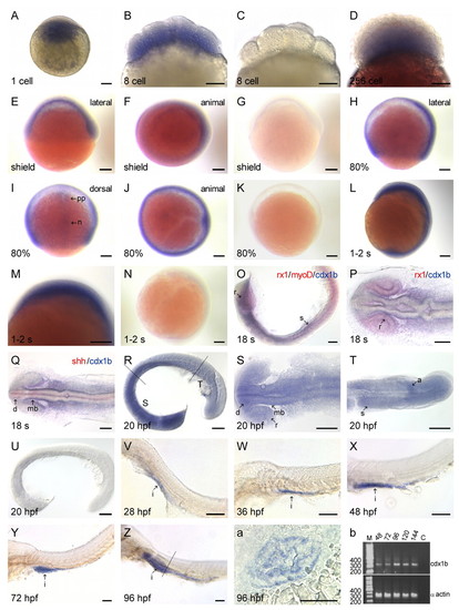
Developmental mRNA expression pattern of the zebrafish cdx1b gene. cdx1b mRNA was detected in one-cell zygote (A), cleavage (B), blastula (D), shield (E,F), 80% epiboly (H-J), 1-2s (L,M), 18s (O-Q), 20 hpf (R-T), 28 hpf (V), 36 hpf (W), 48 hpf (X), 72 hpf (Y) and 96 hpf (Z) stages. (C,G,K,N,U) Embryos hybridized with the sense RNA probe. Double-labeled in situ hybridization showed the expression of cdx1b compared with either rx1, myoD (O,P) or shh (Q). The two lines in R show different boundaries of a 20 hpf embryo viewed at higher magnification in S and T. (a) Cryostat transverse section along the plane of the line shown in Z. (b) Semiquantitative RT-PCR revealed cdx1b expression levels in embryos from different developmental stages. a, anus; d, diencephalon; i, intestine; mb, midbrain; n, notochord; pp, prechordal plate; r, retina; s, somite. Scale bars: 100 μm. |

cdx1b antisense MO knockdown analyses. cdx1b 24 hpf (A-F) and 48 hpf (M,N) zebrafish morphants; cdx1b-4mm-MO-injected 24 hpf (G-I) and 48 hpf (O,P) embryos; wild-type 24 hpf (J-L) and 48 hpf (Q,R) embryos. Green fluorescence was not detected in cdx1b MO and CMV-cdx1b-mo-GFP co-injected (T) 27 hpf embryos, whereas bright green fluorescence was detected in cdx1b-4mm MO and CMV-cdx1b-mo-GFP co-injected embryos (S). The expression patterns of shh were compared in 28 hpf wild type (U,V) and morphants (W,X). Scale bars: 100 μm. a, atrium; bp, basal plate midbrain; fp, floor plate; hy, hypothalamus; v, ventricle; wt, wild type; zli, zona limitans intrathalamica. |

Inhibition of zebrafish cdx1b function affects expression of some digestive organ marker genes and causes hypoplastic growth of the liver, pancreas and intestines. Seventy-two hpf (A,D) and 54 hpf (G) wild type and 72 hpf (B,C,E,F) and 54 hpf (H) morphants were respectively labeled with lfabp/ifabp (A-C), trypsin (D-F) and insulin (G,H) probes. Real-time quantitative PCR (I) indicated a reduction in expression levels of different marker genes in 72 hpf morphants. Histological analyses of paraffin transverse (J-L) and sagittal (S-U) sections of 96 hpf wild-type and transverse (M-R) and sagittal (V-X) sections of morphants are shown. The inset in K indicates the sectioning planes on digestive tracts shown in J-R. Mid-intestine and posterior-intestine regions of wild-type (T,U) and morphant (W,X) at a higher magnification are shown. Scale bars: 100 μm. a, anus; es, esophagus; ep, exocrine pancreas; i, intestine; l, liver; wt, wild type. EXPRESSION / LABELING:
|
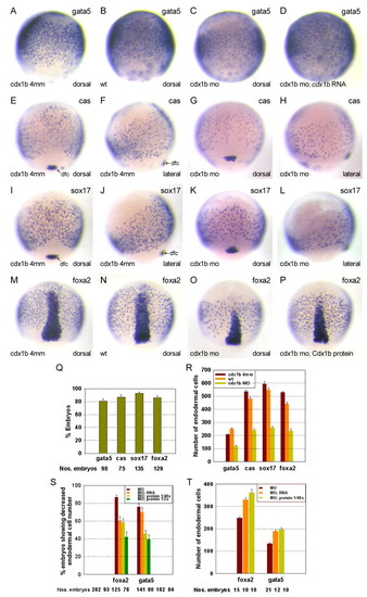
Analyses of relationships between cdx1b and downstream factors of Nodal signaling in zebrafish. Reductions in respective gata5-, cas-, sox17- and foxa2-expressing endodermal cell numbers were observed in 85% epiboly morphants (C,G,H,K,L,O) compared with respective cdx1b-4mm-MO-injected (A,E,F,I,J,M) and wild-type (B,N) embryos. Embryos co-injected with either cdx1b mRNA or protein showed restoration of respective gata5- (D) and foxa2-expressing (P) endodermal cell numbers. Percentages of morphants showing decreases in the respective numbers of gata5-, cas-, sox17- and foxa2-expressing endodermal cells (Q). Comparison of the respective gata5-, cas-, sox17- and foxa2-expressing endodermal cell numbers in morphants (yellow bars), and wild type (orange bars) and cdx1b-4mm-MO-injected (brown bars) embryos (n=10 each) (R). Comparison of percentages of embryos showing decreased foxa2- or gata5-expressing endodermal cell numbers in respective embryos injected with either cdx1b MO (brown bars), cdx1b MO and cdx1b mRNA (orange bars), or cdx1b MO and different amounts of Cdx1b protein (1/40x, yellow bars; 1/2x, green bars) (S). Comparison of the respective foxa2- and gata5-expressing endodermal cell numbers in morphants (brown bars) and embryos co-injected with cdx1b MO and either cdx1b mRNA (orange bars) or protein (yellow bars) (T). Error bars represent standard error. Dfc, dorsal forerunner cell; wt, wild type. EXPRESSION / LABELING:
PHENOTYPE:
|

Zebrafish cdx1b epiboly morphant showed no ectoderm or mesoderm defects. Expression of fgf3 in wild type (A) and bud morphants (B). gsc expression in wild type (C) and shield morphants (D). ntl expression in 80% morphants (F) and wild-type (E) embryos. fb, forebrain; n, notochord; pp, prechordal plate; r4, rhombomere 4; wt, wild type. Scale bars: 100 μm. |
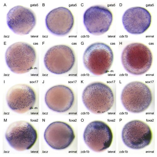
Effects of ectopic cdx1b expression on respective gata5-, cas-, sox17- and foxa2-expressing endodermal cell numbers in zebrafish. Increases in the numbers of gata5- (C,D), cas- (G,H), sox17- (K,L) and foxa2-expressing (O,P) endodermal cells were detected in 85% epiboly embryos ectopically expressing cdx1b when compared with lacZ (A,B,E,F,I,J,M,N) ectopically expressing epiboly embryos. Scale bars: 100 μm. Dfc, dorsal forerunner cell. |
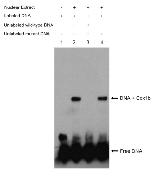
Electrophoretic mobility shift assay of the Cdx1b protein. The biotin-labeled wild-type oligonucleotide was mixed with buffer (lane 1) or 10 μg of nuclear extract prepared from COS-1 cells transfected with pcDNA3-cdx1b-Myc-His plasmid (lanes 2-4). Binding was completely abolished by the addition of an unlabeled wild-type oligonucleotide competitor in a 50-fold molar excess (lane 3), whereas specific binding was maintained when the same amount of the excess mutant oligonucleotide competitor was added (lane 4). |
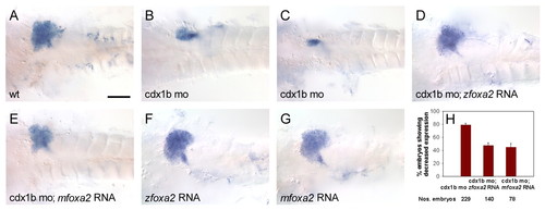
Injection of either zebrafish or mouse Foxa2 mRNA restored the expression domain of ceruloplasmin in the liver of cdx1b morphants. Expression of ceruloplasmin in a 53 hpf wild type (A), respective morphants (B,C), an embryo co-injected with zebrafish foxa2 mRNA and cdx1b MO (D), an embryo co-injected with mouse Foxa2 mRNA and cdx1b MO (E), and respective embryos injected with either zebrafish foxa2 (F) or mouse Foxa2 (G) mRNA alone. (H) Comparison of the percentages of embryos showing reduced levels of the ceruloplasmin expression domain in respective embryos injected with either the cdx1b MO, zebrafish foxa2 mRNA and cdx1b MO, or mouse Foxa2 mRNA and cdx1b MO. Scale bar: 100 μm. wt, wild type. EXPRESSION / LABELING:
|
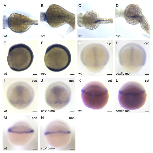
Analyses of relationships between cdx1b and components of Nodal signaling. A slightly increased expression area of cdx1b in the intestines was detected in a 48-hpf squintcz35 mutant embryo (n=9) (B) when compared with a sibling wild-type embryo (A). However, a subsequent semiquantitative RT-PCR revealed no significant differences when comparing cdx1b expression levels in 48-hpf squintcz35 homozygous mutants with their sibling wild-type or AB wild-type embryos. No differences in cdx1b expression was observed in 48-hpf cycb16 (n=7) (D) and 4-6s oepm134 mutants (n=26) (F) compared with sibling wild-type embryos (C,E). Respective expression levels of cyclops (n=54) and oep (n=54) in 85% epiboly cdx1b morphants (H,J) were not altered compared with wild-type embryos (G,I). Similar expression levels of sqt (n=172) and bon (n=91) were detected in 30% (L) and 40% (N) G,I). Similar expression levepiboly morphants when compared with respective wild-type embryos (K,M EXPRESSION / LABELING:
|
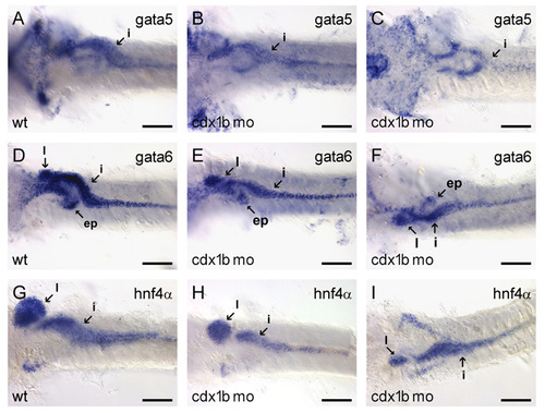
Inhibition of cdx1b function affects expression domains of respective gata5, gata6 and hnf4α in the liver, pancreas and intestines of cdx1b morphants. Expression of gata5 in 54-hpf wild-type embryos (A) and respective morphants (B,C); gata6 expression in wild-type embryos (D) and respective morphants (E,F); hnf4α expression in wild-type embryos (G) and respective morphants (H,I). Ep, exocrine pancreas; i, intestine; l, liver. Scale bars: 100 μm. |

Unillustrated author statements EXPRESSION / LABELING:
|