
- Title
-
Otx1l, Otx2 and Irx1b establish and position the ZLI in the diencephalon
- Authors
- Scholpp, S., Foucher, I., Staudt, N., Peukert, D., Lumsden, A., and Houart, C.
- Source
- Full text @ Development
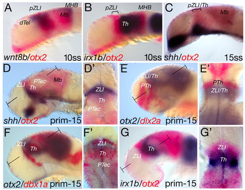
Mapping of otx2 expression relative to markers of the MDT. Whole-mount double in situ-hybridised wild-type embryos are mounted with anterior to the left and dorsal up. The markers and stages are indicated. (A) wnt8b marks the pZLI territory and shares the same anterior border as otx2 at ten somites (14 hpf). (B) At the same stage, otx2 is expressed in the territory from the pZLI to the midbrain, whereas irx1b and otx2 are co-expressed in the presumptive thalamic area and anterior midbrain. (C,D) At 15 somites (16.5 hpf), shha expression is induced within the otx2-positive domain (C) and its anterior border coincides with the anterior expression border of otx2 at prim-15 (30 hpf; D). (D') A cross-section shows that the otx2 expression domain marks the ZLI and the thalamus whereas the ZLI is only shha positive. In the pretectum, otx2 is weakly expressed. (E,E') dlx2a marks the territory of the prethalamus and is anteriorly adjacent to the otx2 expression domain (E); the cross-section reveals the abutting expression domains (E'). (F,F') Similarly to the shha expression domain, the expression domain of dbx1a overlaps with that of otx2 at the anterior ZLI domain and at the thalamus. (G,G') At 30 hours, the expression domain of irx1b persists in the thalamus, whereas the ZLI domain is only otx2 positive. T-shaped brackets mark the plane of the section in the following picture. dTel, dorsal telencephalon; Mb, midbrain; MHB, midbrain-hindbrain boundary; prim-15, primordium stage 15; PTec, pretectum; PTh, prethalamus; pZLI, presumptive zona limitans intrathalamica; ss, somite stage; Th, thalamus; ZLI, zona limitans intrathalamica. EXPRESSION / LABELING:
|
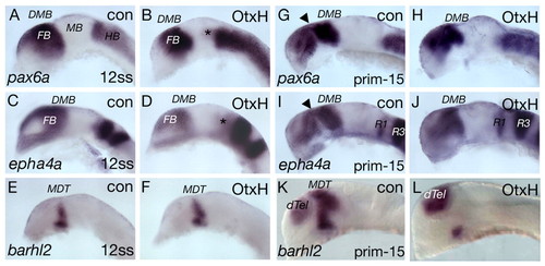
Forebrain marker gene expression is unaltered in Otx1l/2 hypomorphic (OtxH) embryos at early stages. Gene expression profile of OtxH embryos at 12 somites (15 hpf; A-F) and prim-15 (30 hpf; G-L). (A,C) pax6a and epha4a mark the forebrain anlage and demarcate at the diencephalic-mesencephalic boundary. (B,D) Although the hindbrain expression domains start to shift anteriorly (asterisks), the forebrain expression domains of these genes are unaltered in OtxH embryos at this stage. (G-J) At prim-15, in control siblings, pax6a and epha4a become downregulated in the mid-diencephalon (marked by arrowheads); this structure is not detectable in OtxH embryos (pax6a: 26/32; epha4a: 36/41). Compared with control siblings, the expression domains of pax6a and epha4a shrink in the anteroposterior direction. (E,F) barhl2 is expressed in the MDT at 12 ss (E) and its expression is indistinguishable from OtxH embryos at the same stage (F). (K) At prim-15, barhl2 marks the dorsal telencephalon and the MDT, including the ZLI (12/30). (L) In OtxH embryos, the ZLI expression is absent, whereas the dorsal telencephalic domain persists. con, control; dTel, dorsal telencephalon; DMB, diencephalic-mesencephalic boundary; FB, forebrain; HB, hindbrain; MB, midbrain; MDT, mid-diencephalic territory; prim-15, primordium stage 15; R1, rhombomere 1; R3, rhombomere 3; ss, somite stage. EXPRESSION / LABELING:
|
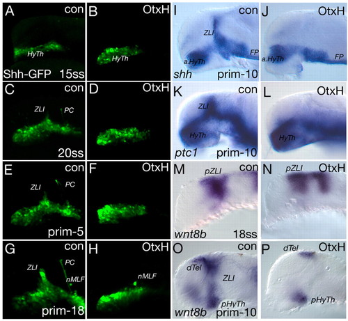
Analysis of the ZLI in OtxH embryos. Confocal microscopy analysis of transgenic shh-GFP OtxH embryos and controls (con) (A-H) and gene expression analysis of OtxH embryos (I-P); the markers and stages are indicated. (A,B) Shh-GFP is detectable in the hypothalamic primordium (HyTh) in control embryos as well as in OtxH embryos at 17.5 hpf. (C-H) From 21 to 32 hpf, the ZLI develops from ventral to dorsal and the posterior commissure (PC) forms at the diencephalic-mesencephalic boundary in control embryos (C,E,G), whereas, in OtxH embryos, neither the ZLI nor the PC is detectable (D,F,H). Interestingly, the nucleus of the medio-longitudinal fascicle (nMLF) is visible in control embryos as well as in OtxH embryos (G,H). (I,J) At prim-10 (28 hpf), the Shh expression domains of the basal plate are unaltered in the anterior hypothalamus (aHyTh) and floor plate (FP) in OtxH embryos, whereas the ZLI, located in the alar plate, is missing compared with control embryos (42/51). (K,L) Similarly, the expression domain of ptc1 is absent at the ZLI in OtxH embryos (31/48). (M,N) Expression of the marker of the presumptive ZLI (pZLI), wnt8b, is unaltered in the diencephalic expression domain at 18 somites (18 hpf), although the MHB expression shifts anteriorly. (O,P) At prim-10, wnt8b is absent in the ZLI (10/12) but persists in the dorsal telencephalon (dTel) and posterior hypothalamus (pHyTh). prim, primordium stage; ZLI, zona limitans intrathalamica. EXPRESSION / LABELING:
|
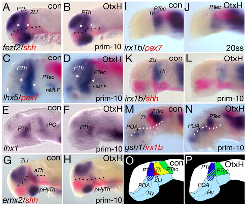
Analysis of the MDT in OtxH embryos. (A-H) Single and double in situ hybridisation of OtxH embryos; the markers and stages are indicated. Shh expression is absent in the ZLI territory in OtxH embryos (B,H,L). Compared to control (con) siblings, markers for the prethalamic anlage - fezl, lhx5 and lhx1 - are still expressed in OtxH embryos and show a slight broadening in the ventral part of the anlage (lhx5: 11/15; fezl (fezf2): 12/20; A-F, marked by arrowheads). In OtxH embryos, the nuclei of the medio-longitudinal fascile is detectable by lhx5 (C,D), whereas the lhx1-positive nuclei of the PC are absent (E,F). emx2 expression in the anterior thalamus (aTh) is absent in OtxH embryos (23/30). (I-N) Prior to ZLI formation, at 20 somites (19 hpf), the irx1b expression domain is absent in OtxH embryos (I,J) and is still absent at prim-10 (28 hours; 24/60; K-N). (A-H) Dotted line marks the border between alar and basal plate. (C,D,M,N) pax7 is expressed in the dorso-lateral part of the pretectum (C,D), whereas gsh1 marks the ventro-medial domain of the pretectum (M,N). (C,D) In OtxH embryos, the pax7-positive part of the pretectum shifts anteriorly and a new border between the lhx5-positive prethalamus and the pax7-positive pretectum is observed (11/15). (M,N) Comparison of the gsh1-positive part of the pretectum in OtxH embryos and their wild-type siblings shows that the pretectum area abuts the gsh1-positive preoptic area (8/14). (O,P) Summary of the observations in OtxH embryos. In wild-type embryos, the MDT is organised in the following way: the lateral halves of the prethalamus (blue), the more medial ZLI (red), the thalamus (yellow) and the pretectum (green). In OtxH embryos, we still find the lateral halves of the prethalamus (blue) and the pretectum (green). Striped areas show the lateral to medial organisation of the area and not an overlap of expression domains. aTh, anterior thalamus; Hy, hypothalamus; nMLF, nucleus of the medio-longitudinal fascile; nPC, nucleus of the posterior commissure; pHyTh, posterior hypothalamus; POA, post-optic area; prim-10, primordium stage 10; PTec, pretectum; PTh, prethalamus; ss, somite stage; Th, thalamus; ZLI, zona limitans intrathalamica. EXPRESSION / LABELING:
|
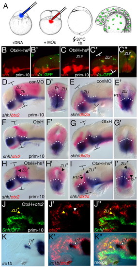
Otx2 rescues the ZLI in OtxH embryos. Gene expression analysis of embryos at prim-10 (28 hpf); the markers are indicated on each panel. (A) Embryos were injected with 60 pg of the heat-shock-inducible otx2-GFP DNA construct into the one-cell-stage blastomere. Embryos were kept at 28°C until reaching the four-cell stage. Then, the double morpholino (MO) mix was injected to block transcription of endogenous otx1l and otx2. At the one-somite stage, the embryos were heat-shocked for 1 hour at 37°C and were fixed at the prim-12 stage. To reliably localise the Otx2-GFP-positive clone, an FITC-coupled antibody staining against GFP and a Fast Red single in situ hybridisation for shh were performed. Injection of otx2-GFP DNA and heat-shock activation led to a random induction of GFP-positive clones in the OtxH embryos. Confocal microscopy analysis of the embryos showed that ectopic induction of shh expression is generally not observed in otx2-GFP clones (B,B′). However, when these clones were located within the MDT, shh expression was induced in these cells (C,C′ and cross-section in C″, 13/16-4/6 detected via &alpha-GFP staining and 9/10 via GFP-ISH). (D,D′) To mark the prethalamic-thalamic boundary, a double in situ hybridisation of otx2 and shh was performed. (E,E′) To mark the prethalamus, a double staining with dlx2a and shh was performed. (F-G′) In control (con) embryos, shh expression is absent at the ZLI. In addition, the otx2 expression domain is smaller (F,F′) and the dlx2a domain is absent (G,G′). By contrast, in embryos in which otx2-GFP is heat-activated, shh expression can be rescued in the presumptive MDT (H,H′), and a cross-section reveals that the induction is independent of the basal shh expression domain (H′). The rescue of shh in small clones is sufficient to induce the expression of dlx2a (5/10; I); for example, its expression was induced uni-laterally in the neural tube (I′). T-shaped brackets mark the plane of the section in the following picture. The dotted line marks the border between the alar and basal plate. (J-K″) otx2 mRNA (150 pg; + red-lineage tracer) was injected into one of 64 cells of OtxH shh-GFP embryos. At 32 hpf, embryos were fixed and stained for GFP (green). The provision of an ectopic otx2 message is able to rescue Shh-GFP expression within the ZLI in OtxH embryos (J-J″, yellow arrowhead), as shown by a co-localisation of the red-lineage tracer of the Otx2+ cells with the green GFP expression. The irx1b expression profile was assessed in the same embryo (K-K″). When Otx2+ cells are located within the presumptive thalamic territory, they express irx1b (white arrowhead), shown by co-localisation of the red lineage tracer with irx1b detection (blue). (K″) Merged picture of Shh-GFP expression, the red lineage tracer and irx1b expression. prim-10, primordium stage 10; PTh, prethalamus; PTh*, rescued prethalamus; Th, thalamus; ZLI, zona limitans intrathalamica; ZLI*, rescued zona limitans intrathalamica. |
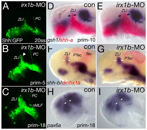
Irx1b sets the posterior boundary of the ZLI. Confocal microscopy analysis of transgenic shh-GFP; irx1b-morpholino (MO) embryos (A-C) and in situ-hybridisation analysis of irx1b-MO embryos (D-I); markers and stages are indicated. (A) irx1b-MO-injected embryos resemble control (con) embryos at the same stage (compare with Fig. 3). (B) At 26 hpf, cells located posterior to the ZLI start to express Shh-GFP ectopically (arrowhead), whereas the posterior commissure (PC) and the nucleus of the medio-longitudinal fascicle (nMLF) form normally. (C) At 32 hpf, the Shh-GFP expression at the ZLI is broadened compared with control-injected embryos (see Fig. 3). (D,E) At prim-10, the Shh expression domain at the ZLI is broadened at the expense of the thalamus (arrows). (F,G) Similarly, the expression domain of shhb expands posteriorly in irx1b morphants (arrows). (D-G) Interestingly, the expression of markers of the pretectum, such as gsh1 and dmbx1a, is unaltered. (H,I) The broadening of the ZLI is also visible by an in situ hybridisation for pax6a, revealing a much wider gap between the prethalamus and the remaining thalamic-pretectal tissue at prim-18 (32 hpf; arrows). nMLF, nucleus of the medio-longitudinal fascicle; PC, posterior commissure; prim, primordium stage; PTec, pretectum; Tec, tectum; ZLI, zona limitans intrathalamica. |