
- Title
-
Regulation and function of Dbx genes in the zebrafish spinal cord
- Authors
- Gribble, S.L., Nikolaus, O.B., and Dorsky, R.I.
- Source
- Full text @ Dev. Dyn.
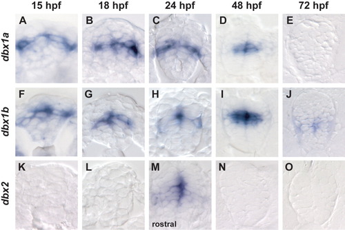
Expression of dbx1a/1b/2 during spinal cord development. Spinal cord cross-sections are shown at five different stages of development for dbx1a, dbx1b, and dbx2. A-E: dbx1a expression is present at 15 hours postfertilization (hpf) though 48 hpf and is absent at 72 hpf. The territory that dbx1a occupies does not change appreciably throughout time, although at 48 hpf (D) dbx1a is no longer expressed in postmitotic neurons. F-J: dbx1b expression is very similar to dbx1a expression, with the exception that it is present at low levels at 72 hpf. K-O: dbx2 expression differs from dbx1a/1b. It is not expressed in the spinal cord until 24 hpf and only in rostral segments at that time and is down-regulated by 48 hpf. EXPRESSION / LABELING:
|
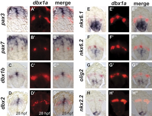
Relationship of dbx1a to other spinal cord patterning genes. A-H″: Spinal cord cross-sections showing double in situ hybridization at 24 hours postfertilization (hpf; dbx2 28 hpf). In each panel patterning genes are in blue (A-H), dbx1a is shown in red (A′-H′), and merged images are shown in (A″-H″). dbx1a expression does not overlap with expression of pax3 (A-A″), pax7 (B-B″), nkx6.1 (E-E″), nkx6.2 (F-F<″), olig2 (G-G″), or nkx2.2 (H-H″). dbx1a completely overlaps with dbx1b (C-C″) and is expressed within the domain of dbx2 (D-D″). |
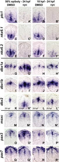
Analysis of patterning defects in cyclopamine-treated embryos. Spinal cord cross-sections of embryos treated with dimethyl sulfoxide (DMSO) or cyclopamine at 50% epiboly or 18 hours postfertilization (hpf) are shown. A-F: The 100 μM cyclopamine blocked expression of ptc1 (A-B″), nkx6.1 (C-D″), and nkx6.2 (E-F″) under both treatment conditions. G,I,K: Expression of dbx1a (G, G″), dbx1b (I, I″), and dbx2 (K, K″) expands ventrally after cyclopamine treatments beginning at 50% epiboly compared with DMSO controls. H,J,L: Similarly, after cyclopamine treatments beginning at 18 hpf, dbx1a (H,H″), dbx1b (J,J″), and dbx2 (L,L″) expand slightly ventrally. M-P: The expression of msxc (M-N″) is unaffected at either stage of treatment, whereas pax3 (O,O″) expression expands ventrally into the dbx1a/1b domain when treated at 50% epiboly, but no expansion is observed in the 18-24 hpf treatment (P,P″). Q,R: In contrast, pax7 (Q-R″) expression does not expand ventrally in either treatment. EXPRESSION / LABELING:
|
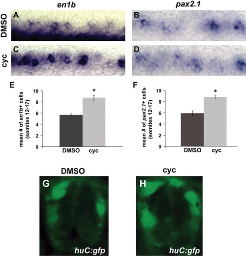
en1b+ and evx1+ interneurons increase in cyclopamine-treated embryos. A-D: Lateral whole-mount views of subtype-specific postmitotic marker are shown with rostral to the left at 24 hours postfertilization (hpf). A-F: en1b+ (A,C,E) and pax2.1+ (B,D,F) interneurons are increased in cyclopamine-treated embryos. Error bars represent SEM. G,H: Representative sections of green fluorescent protein (GFP) expression in Tg(elavl3:EGFP)zf8 embryos are shown. There is no change in the number of Hu-positive neurons after cyclopamine treatment. |
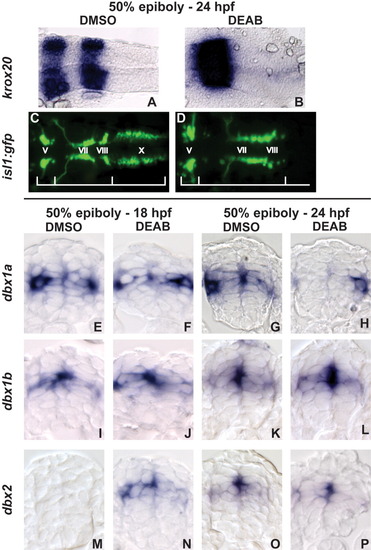
Analysis patterning defects in 4-(diethylamino)-benzaldehyde (DEAB) -treated embryos. A,B: Dorsal view of 24 hours postfertilization (hpf) embryos showing krox20 mRNA expression. Dimethyl sulfoxide (DMSO) -treated embryos show krox20 expression in rhombomeres 3 and 5 and DEAB-treated embryos only show expression in rhombomere 3. C,D: dorsal view of green fluorescent protein (GFP) expression in cranial nuclei in Tg(isl1:GFP)rw0 embryos at 24 hpf. DMSO-treated embryos have normal GFP expression, and DEAB-treated embryos have loss of the vagal (X) nuclei and abnormal positioning of the VIIIth cranial nuclei. E-P: Spinal cord cross-sections of embryos treated with DMSO or DEAB. dbx1a mRNA expression is unaffected when embryos are fixed at 18 hpf (E,F) but by 24 hpf, progenitor expression and often postmitotic neuron expression of dbx1a is lost (G,H). dbx1b expression is unaffected at 18 hpf (I,J) and 24 hpf (K,L) in DEAB-treated embryos. dbx2 expression is prematurely induced at 18 hpf in DEAB-treated embryos (M,N) and is maintained at 24 hpf (O,P). EXPRESSION / LABELING:
PHENOTYPE:
|
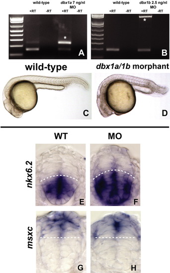
dbx1a and dbx1b morpholinos cause spinal cord patterning defects. A,B: RT-PCR for dbx1a (A) and dbx1b (B) in morphants show unspliced products (asterisk in each gel) at a concentration of 7 ng/nl for dbx1a and 2.5 ng/nl for dbx1b. Both morpholinos were injected simultaneously to produce dbx1a/1b loss-of-function embryos. C,D: Whole-mount views of a wild-type (C) and dbx1a/1b morphant (D) embryo at 24 hours postfertilization (hpf) with rostral to the left. dbx1a/1b morphants have a shortened rostral/caudal axis and hindbrain morphology defects. E-H: Spinal cord cross-sections of wild-type and dbx1a/1b morphants at 24 hpf. E,F: The nkx6.2 expression is expanded dorsally in dbx1a/1b morphants compared with wild-type. G,H: The msxc expression is unaffected in dbx1a/1b morphants. |
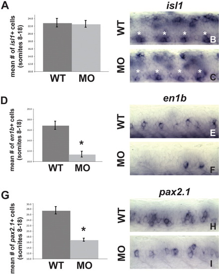
dbx1a/1b knockdown causes postmitotic neuron defects. Postmitotic neuron analysis of dbx1a/1b morphants. Lateral whole-mount views of subtype-specific postmitotic marker are shown with rostral to the left at 24 hours postfertilization (hpf). A-C: isl+ motoneurons are unaffected in dbx1a/1b morphants. D-I: en1b+ (D-F) and pax2.1+ (G-I) interneurons are decreased in dbx1a/1b morphants. Error bars represent SEM. |