
- Title
-
Expression of zebrafish ROR alpha gene in cerebellar-like structures
- Authors
- Katsuyama, Y., Oomiya, Y., Dekimoto, H., Motooka, E., Takano, A., Kikkawa, S., Hibi, M., and Terashima, T.
- Source
- Full text @ Dev. Dyn.
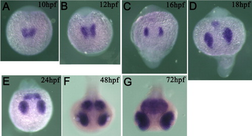
Expression pattern of rora1 detected by whole-mount in situ hybridization. A-D: Dorsal views of developing brain. E-G: Views from the anterior top. Embryonic expression of rora1 starts in the eye rudiments from the 1-somite stage (10 hours postfertilization [hpf]), and expression in the presumptive tectum was detected from the 14-somite stage (16 hpf). EXPRESSION / LABELING:
|
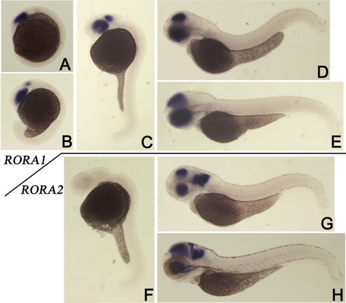
A-H: Lateral views of in situ hybridization specimens in which expression of rora1 (A-E) or rora2 (F-H) was detected. A: At the five-somite stage (12 hours postfertilization [hpf]). B: At the 18-somite stage (18 hpf). C,F: Expression of rora1 (C) was observed in the eyes and midbrain, but expression of rora2 (F) was not detected in 24 hpf specimens. D,G: Expression of rora1 (D) and rora2 (G) was similarly observed in the eyes and tectum, but only rora2 expression was observed in the hindbrain of 2 days postfertilization (dpf) specimens (G). H: Expression of rora2 in the cerebellum is observed in 3 dpf specimens. EXPRESSION / LABELING:
|
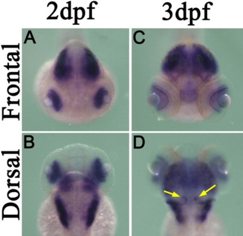
Expression pattern of rora2 in the larval brain and eyes detected by whole-mount in situ hybridization. A-D: Frontal (A,C) and dorsal (B,D) views of 2 days postfertilization (dpf; A,B) and 3 dpf (C,D) specimens are shown. Frontal and dorsal views of a 3 dpf larva clearly show rora2 expression in the eyes and differentiating Purkinje cells (indicated by arrows in D), respectively. EXPRESSION / LABELING:
|
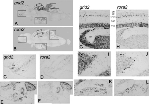
Expression pattern of grid2, glutamate receptor delta2 (A,C,E,G,I,K) and rora2 (B,D,F,H,J,L) was detected by in situ hybridization in adjacent sections of an adult brain of zebrafish. High magnification views of the boxed regions in A and B are shown in the ventral part of the telencephalon (C,D), the ventral part of the midbrain (E,F), the optic tectum and anterior part of the cerebellum (G,H), the posterior part of the cerebellum (I,J), and the dorsal part of the pons (K,L). Colocalized expression of these two genes was observed in the cerebellar-like structures. Abbreviations are the same as used in Wullimann et al. ([1996]). Vdm, dorsal nucleus of V; Vv, ventral nucleus of V; PTN, posterior tuberal nucleus; Hc, caudal zone of periventricular hypothalamus; CM, corpus mamillare; SM, stratum marginale; SO, stratum opticum; SFGS, Stratum fibrosum et griseum superficiale; SGC, stratum griseum centrale; PGZ, periventricular gray zone; Val, lateral division of valvula cerebelli; CCe, corpus cerebelli; PL, Purkinje cell layer; LCa, lobus caudalis cerebelli; MON, medial octavolateralis nucleus. EXPRESSION / LABELING:
|
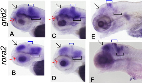
Developmental expression pattern of grid2 (A,C,E) and rora2 (B,D,F) in 48 hpf (A,C), 72 hpf (B,D), and 120 hpf (E,F) specimens. Expression of both genes was observed in the telencephalon (red arrows), the optic tectum (black arrows), upper rhombic lip (the cerebellum, blue boxes), and the lower rhombic lip (black boxes) and eyes. Expression of grid2 in the cerebellum is wider than that of rora2 along the anterior-posterior axis. In C, nonspecific staining was observed in surface of the cranial cavity. |