
- Title
-
The bHLH transcription factor hand2 is essential for noradrenergic differentiation of sympathetic neurons
- Authors
- Lucas, M.E., Muller, F., Rudiger, R., Henion, P.D., and Rohrer, H.
- Source
- Full text @ Development
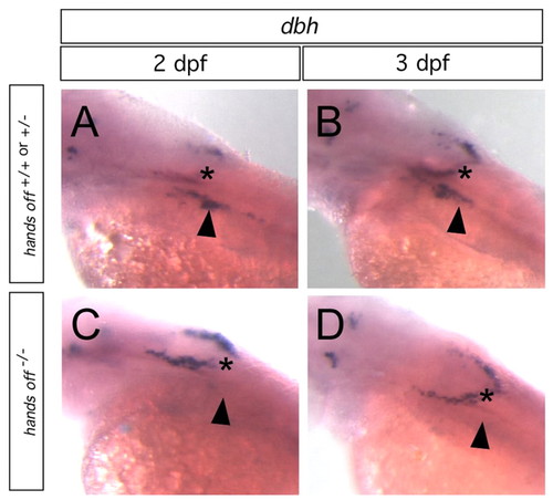
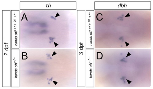
The development of dbh-positive cervical sympathetic ganglia (CSG) is severely affected in hands off embryos. Whole-mount in situ hybridization for dbh in wild-type (A,B) and hands off mutant embryos C,D) at 2 dpf (A,C) and 3 dpf (B,D). dbh-positive CSG cells (arrowheads) are missing in hands off embryos, whereas dbh-positive cells in the dorsally located medulla oblongata are not affected [asterisks; noradrenergic neurons of the medulla oblongata/area postrema form a semicircle (Holzschuh et al., 2003), giving the impression of two stripes in the oblique view from lateral shown]. EXPRESSION / LABELING:
PHENOTYPE:
|
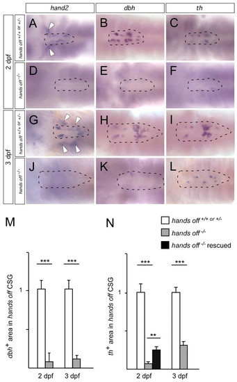
dbh and th expression in cervical sympathetic ganglia of wild type and hands off mutants. Whole-mount in situ hybridizations for hand2 (A,D,G,J), dbh (B,E,H,K) and th (C,F,I,L) viewed from dorsal after dissection of the yolk sac and spinal cord. hand2 labels sympathetic ganglia (indicated by dashed circle) and more ventrally located enteric neurons (diffuse staining below the focal plane; white arrowheads) that migrate to a more lateral position at 3 dpf (G). hands off embryos at 2 dpf (D) and 3 dpf (J) are devoid of hand2 expression and display only very few dbh+ (E,K) and th+ cells (F,L). (M) Area of dbh-expressing cells at 2 and 3 dpf in hands off mutants compared with wild type. (Number of embryos analysed: nine wild type, three mutant for 2 dpf; 11 wild type, seven mutant for 3 dpf.) (N) Area of th-expressing cells at 2 and 3 dpf in hands off mutants compared with wild type. Data are presented as mean ± s.e.m. (Number of embryos analysed: 28 wild type, ten mutant for 2 dpf; 41 wild type, 16 mutant for 3 dpf.) The injection of wild-type hand2 mRNA resulted in a significant increase in the number of th-expressing cells in hands off mutants analysed at 2 dpf (n=10). (**P<0.01; ***P<0.001.) EXPRESSION / LABELING:
|
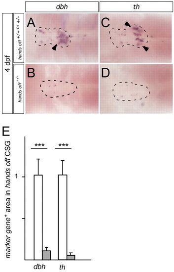
dbh and th expression in 4 dpf cervical sympathetic ganglia of wild type and hands off mutants. Whole-mount in situ hybridizations for dbh (A,B) and th (C,D) as in Fig. 2. dbh and th label sympathetic ganglia. In addition, in the caudal region of the CSG a cluster of more ventrally located chromaffin cells with more intensive staining becomes apparent at 4 dpf (arrowheads). hands off embryos show only very few dbh+ (C) and th+ cells (D), demonstrating that noradrenergic differentiation is prevented in 4 dpf CSG neurons and chromaffin cells. (E) Area of total dbh- and th-expressing cells in hands off mutants compared with wild type. Data are presented as mean ± s.e.m. (Number of embryos analysed: 14 wild type, six mutant for dbh; 12 wild type, seven mutant for th.) (***P<0.001.) |
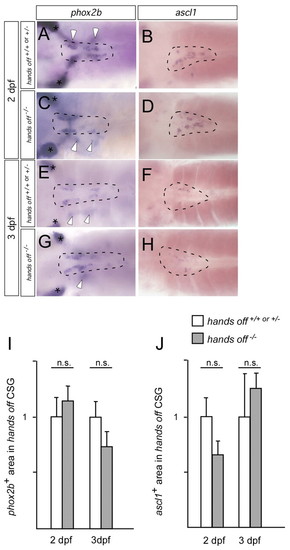
phox2b and ascl1 expression in cervical sympathetic ganglia of wild type and hands off mutants. phox2b and ascl1 whole-mount in situ hybridizations viewed from dorsal. phox2b labels sympathetic ganglia (dashed circle) and more ventrally located enteric neurons (diffuse staining from below focus plane; white arrowheads). The gut was removed from ascl1-stained embryos. Control and hands off embryos show the same expression of phox2b at 2 dpf (A,C) and 3 dpf (E,G). Also ascl1 expression is not affected in hands off embryos at 2 dpf (B,D) and 3 dpf (F,H). The area and intensity of ascl1 expression is, however, strongly reduced at 3 dpf for both wild-type and hands off embryos. (I) Area of phox2b-expressing cells at 2 and 3 dpf in hands off mutants compared with wild type. (Number of embryos analysed: 15 wild type, five mutant for 2 dpf; 12 wild type, four mutant for 3 dpf.) (J) Area of ascl1-expressing cells at 2 and 3 dpf in hands off mutants compared with wild type. (Number of embryos analysed: ten wild type, seven mutant for 2 dpf; four wild type, four mutant for 3 dpf.) Data are presented as mean ± s.e.m.; n.s. not significant. phox2b staining of epibranchial placodes is indicated by asterisks. EXPRESSION / LABELING:
|
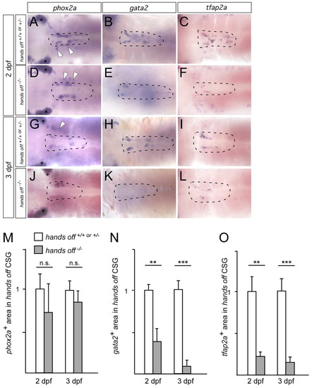
phox2a, gata2 and tfap2a expression in cervical sympathetic ganglia of wild type and hands off mutants. Whole-mount in situ hybridizations viewed from dorsal. (A,D,G,J) phox2a-labeled sympathetic ganglia (dashed circle) and enteric neurons (white arrowheads). Control and hands off embryos show the same expression of phox2a at 2 dpf (A,D) and 3 dpf (G,J). gata2 expression is strongly reduced in hands off embryos (E,K) compared with wild-type embryos (B,H). tfap2a expression is massively reduced in hands off embryos (F,L) compared with wild type (C,I). (M) Area of phox2a-expressing cells at 2 and 3 dpf in hands off mutants compared with wild type. (Number of embryos analysed: seven wild type, three mutant for 2 dpf; ten wild type, five mutant for 3 dpf.) (N) Area of gata2-expressing cells at 2 and 3 dpf in hands off mutants compared with wild type. (Number of embryos analysed: six wild type, five mutant for 2 dpf; 12 wild type, nine mutant for 3 dpf.) (O) Area of tfap2a-expressing cells at 2 and 3 dpf in hands off mutants compared with wild type. (Number of embryos analysed: five wild type, 14 mutant for 2 dpf; 15 wild type, four mutant for 3 dpf.) Data are presented as mean ± s.e.m.; n.s. not significantly different (**P<0.01, ***P<0.001). Phox2a staining of epibranchial placodes is indicated by asterisks. EXPRESSION / LABELING:
|
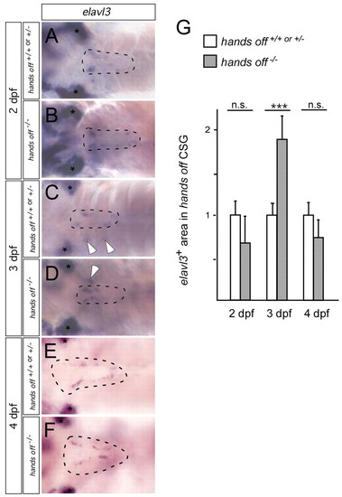
elavl3 (HuC) expression in cervical sympathetic ganglia of wild type and hands off mutants. elavl3 whole-mount in situ hybridizations viewed from dorsal. elavl3-labeled sympathetic ganglia (dashed circle) and enteric ganglia (white arrowhead) are indicated. Expression of elavl3 in control and hands off embryos at 2 dpf (A,B), 3 dpf (C,D) and 4 dpf (E,F). (G) Area of elavl3-expressing cells at 2, 3 and 4 dpf in hands off mutants compared with wild type. (Number of embryos analysed: 13 wild type, six mutant for 2 dpf; 36 wild type, 13 mutant for 3 dpf; nine wild type, seven mutant for 4 dpf). Data are presented as mean ± s.e.m.; n.s. not significantly different; ***P<0.001; asterisks label elavl3 (HuC) staining of epibranchial placodes. |
th and dbh expression in the locus coeruleus of wild type and hands off mutants. th and dbh whole-mount in situ hybridizations viewed from dorsal. th (A,B) and dbh (C,D) expression in the locus coeruleus is not different between wild type (A,C) and hands off mutants (B,D). Two and 3 dpf embryos are shown for th and dbh, respectively. The locus coeruleus is indicated by arrowheads. EXPRESSION / LABELING:
|
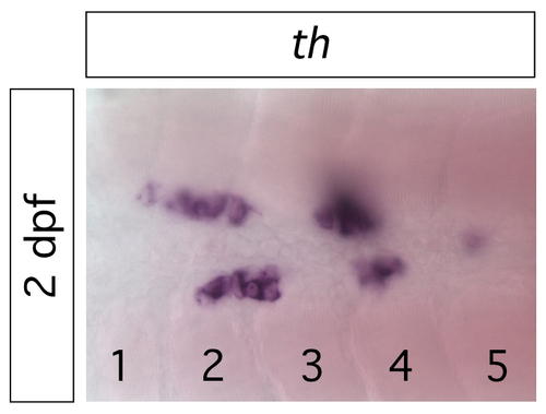
Morphology of cervical sympathetic ganglia in 2 dpf zebrafish embryos. After whole-mount in situ hybridisation for th as a marker for sympathetic neurons, dorsal hindbrain/spinal cord and yolk sac were dissected and viewed from dorsal using 40× objective. Please note individual cells with nuclei spared from the in situ staining reaction in the plane of focus and other cells that give a blurred appearance, as they are out of focus. The embryo shown was selected for the number of cells within one focus plane. Somite numbers are indicated to document that cervical sympathetic ganglion cells are mainly located betwen somites 1 and 4. EXPRESSION / LABELING:
|
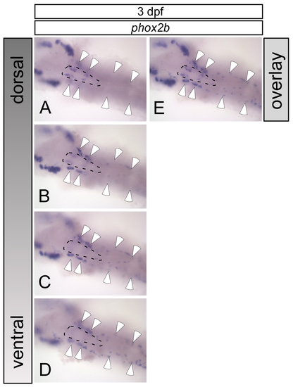
Localisation of cervical sympathetic and enteric neurons in 3 dpf zebrafish embryos. (A-D) Whole-mount in situ hybridisation for phox2b as marker for autonomic cells was carried out. After dissection of dorsal hindbrain/spinal cord and yolk sac, a stack of pictures was made focussing from dorsal (A) to ventral (D) through the embryo. This illustrates the dorsal location of the cervical sympathetic ganglion (dashed circle), restricted to a relatively short anteroposterior area (A). Enteric neurons (white arrowheads) that appear as diffuse staining in A show up in more ventral focal planes (B-D), and extend to more rostral and caudal regions down to the anus (not shown). (E) An optical overlay of images in A to D to illustrate the different lateral positions of enteric and cervical sympathetic ganglion cells. EXPRESSION / LABELING:
|
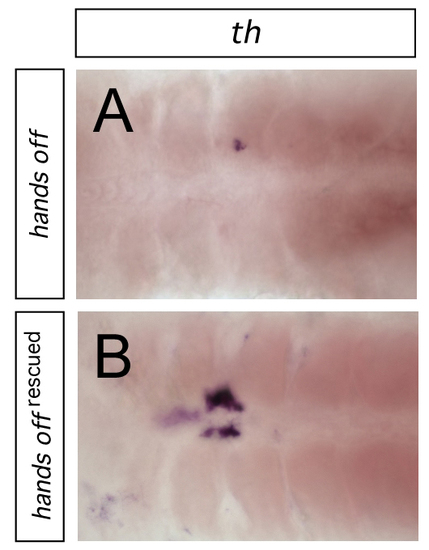
Rescue of th expression in cervical sympathetic ganglia of 2 dpf hands off zebrafish embryos. hands off mutant embryos, (A) without or (B) with injection of synthetic hand2 mRNA, stained for th expression. Rescued hands off mutants displayed a highly significant increase in th-positive cells when compared with uninjected hands off embryos (see Fig. 2N) |

Unillustrated author statements EXPRESSION / LABELING:
|