
- Title
-
Myocyte-specific enhancer factor 2A is essential for zebrafish posterior somite development
- Authors
- Wang, Y., Qian, L., Dong, Y., Jiang, Q., Gui, Y., Zhong, T.P., and Song, H.
- Source
- Full text @ Mech. Dev.
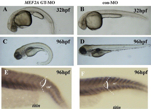
MEF2A morphants display a downward tail curvature and have U-shaped posterior somites although the somite borders are generated normally. (A–F) Lateral views with anterior to the left. The overall morphology of a MEF2A MO-injected (A and C) and control MO-injected (B and D) embryos at 32 hpf (A and B) and 96 hpf (C and D). Note that the MEF2A morphants display a downward tail curvature (A and C) (n = 50 embryos). (E and F) Lateral views of titin expression, which marks each segment border in MEF2A GT-MO-injected (E) and control MO-injected (F) embryos at 96 hpf. Injection of control MO results in chevron-shaped somites with an angle of 95° (n = 20 embryos), while injection of MEF2A GT-MO results in a more obtuse angle of the somite (130°) (n = 19 embryos). |
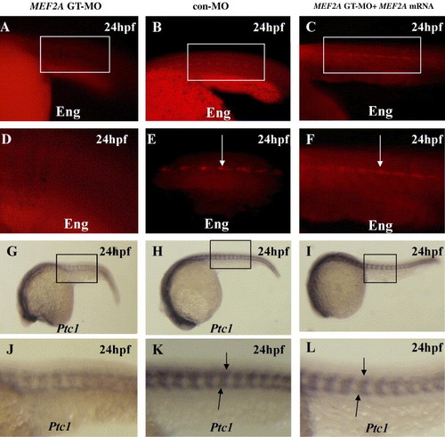
MEF2A morphants exhibit Hedgehog-associated defects in slow muscle. (A–F) Lateral views of somites 8–13 in whole-mount embryos at 24 hpf. Wild-type embryos show strong Engrailed expression in muscle pioneers (arrow) (B and E) (n = 25 embryos). Engrailed expression in MEF2A morphants is mostly absent, though very weak expression can occasionally be observed (A and D) (n = 20 embryos). At 24 hpf, embryos injected at the 1- to 2-cell stage with 50 pg of MEF2A mRNA plus 10 ng of MEF2A GT-MO (C and F) showed rescue of strong Engrailed expression in the muscle pioneers (arrow) (n = 20 embryos). (G–L) Lateral views of whole-mount embryos with the head to the left at 24 hpf. Wild-type embryos exhibit strong expression of ptc1 (H and K) (n = 30 embryos), while MEF2A morphants (G and J) (n = 20 embryos) show weaker levels of ptc1 expression. Also, embryos injected at the 1- to 2-cell stage with 50 pg of MEF2A mRNA plus 10 ng of MEF2A GT-MO (I and L) showed rescue of strong ptc1 expression (arrow) (n = 20 embryos). |
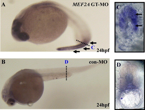
MEF2A inhibition results in induction of apoptosis in the posterior somites. Embryos were fixed at 24 hpf for TUNEL staining. Black arrows indicate widespread apoptosis in the posterior somites (A, compare to B) (A, n = 20 embryos; B, n = 28 embryos). All embryos are shown in lateral view with the head to the left. Cross-sections of MEF2A GT-MO-injected and control MO-injected embryos are shown in C (n = 5 embryos) and D (n = 5 embryos), respectively. A ubiquitous distribution of apoptosis in the muscles could be found through the cross-sections. PHENOTYPE:
|
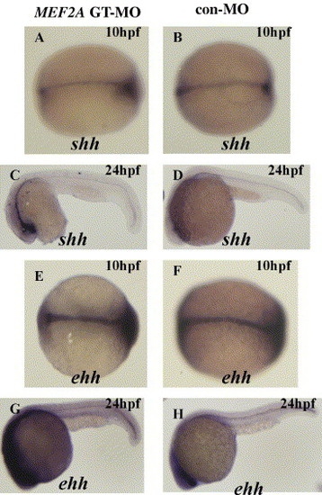
MEF2A is not required for shh and ehh transcription. (A, B, E and F) (A, n = 20 embryos; B, n = 20 embryos; E, n = 20 embryos; F, n = 18 embryos) Dorsal views of whole-mount embryos at the bud stage (10 hpf). (C, D, G and H) (A, n = 20 embryos; B, n = 22 embryos; E, n = 20 embryos; F, n = 20 embryos) Lateral views of whole-mount embryos with the head to the left at 24 hpf. Expression in MEF2A morphants of both shh (A and C) and ehh (E and G) is similar to that observed in control embryos (B, D, F and H). |
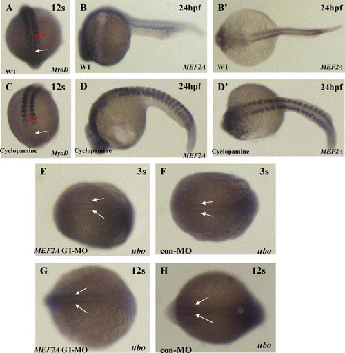
Hedgehog signaling negatively regulates MEF2A expression. (A and C) Dorsal views of whole-mount embryos at the 12-somite stage to show MyoD expression. Wild-type embryos (A, n = 15 embryos) exhibit adaxial MyoD expression throughout the somitic (red arrow) and presomitic (white arrow) mesoderm, while embryos (C, n = 10 embryos) treated with 25 μM cyclopamine at 6 hpf lack expression in the somitic (red arrow) and in the presomitic (white arrow) mesoderm. (B and D) (B, n = 15 embryos; D, n = 10 embryos) Lateral views of whole-mount embryos with the head to the left at 24 hpf. (B′ and D′) (B, n = 15 embryos; D, n = 10 embryos) Dorsal views of whole-mount embryos with the head to the left at 24 hpf. MEF2A expression is increased in the somites of cyclopamine-treated embryos. (E–H) MEF2A is not required for ubo transcription. Dorsal views of whole-mount embryos at the 3-somite stage (E and F) (E, n = 20 embryos; F, n = 20 embryos) and the 12-somite stage (G and H) (G, n = 20 embryos; H, n = 20 embryos) to show ubo expression. (F) At the 3-somite stage, ubo is expressed in the adaxial cells (white arrow). (H) At the 12-somite stage, ubo is expressed prominently in the newly formed posterior somites (white arrow). Expression in MEF2A morphants of ubo is similar to that observed in control embryos (E and G). EXPRESSION / LABELING:
|
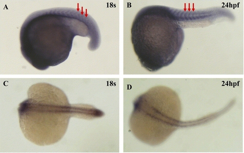
Expression Patterns sf MEF2A during zebrafish embryogenesis. Expression of MEF2A in wild-type embryos at the 18-somite stage (A, C) and at 24 hpf (B, D). MEF2A is expressed in the somites (red arrows). (A, B) Dorsal views of whole-mount embryos with the head to the left. (C, D) Lateral views of whole-mount embryos with the head to the left. EXPRESSION / LABELING:
|
Reprinted from Mechanisms of Development, 123(10), Wang, Y., Qian, L., Dong, Y., Jiang, Q., Gui, Y., Zhong, T.P., and Song, H., Myocyte-specific enhancer factor 2A is essential for zebrafish posterior somite development, 783-791, Copyright (2006) with permission from Elsevier. Full text @ Mech. Dev.