
- Title
-
bmp1 and mini fin are functionally redundant in regulating formation of the zebrafish dorsoventral axis
- Authors
- Jasuja, R., Voss, N., Ge, G., Hoffman, G.G., Lyman-Gingerich, J., Pelegri, F., and Greenspan, D.S.
- Source
- Full text @ Mech. Dev.
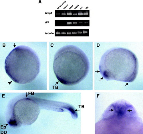
Temporal and spatial distributions of bmp1 expression. (A) RT-PCR analysis of bmp1 and mfn/tll1 RNA levels at different stages of zebrafish development. Expression of tubulin is shown as a control for loading. Each experiment was performed three times with similar results. Spatial bmp1 expression patterns at different stages. (B) Ninety percent epiboly, lateral view, dorsal is to the right. Arrow, bmp1 signal in the ventral quadrant of the animal pole; Arrowhead, signal at the ventral margin. (C) Bud stage, lateral view, dorsal is to the right. TB and arrow denote bmp1 signal in the tail bud. (D) Fifteen somite stage, lateral view, anterior is to the left. Arrows show bmp1 signal in dorsal diencephalon, future tectum, caudal somites/tail bud. (E) Twenty-four hours post fertilization embryo, lateral view, anterior is to the left. Bmp1 signal is detected in pectoral fin bud (FB), Epiphysis (Ep), dorsal diencephalon (DD), and caudal somites/tail bud (TB). (F) Twenty-four hours post fertilization embryo brain, dorsal view, showing bmp1 mRNA localization to the Zona Limitans Interthalamica (ZLI). |
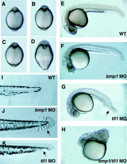
Simultaneous knockdown of both bmp1 and mfn/tll1 expression severely dorsalizes the zebrafish embryo. Morpholinos, both targeted against 5′-UTR sequences and transcription start site, were injected at a concentration of 0.5 mM into 1–2 cell stage embryos, to a final concentration of 0.5 μM in the embryo. Embryos are shown at 12 hpf (dorsal views) (A–D) or 24 hpf (E–H) either uninjected (A and E), or injected with bmp1 (B and F) or tll1 (C and G) morpholinos, or with morpholinos to both genes (D and H). Tails are shown at 55 hpf (I–K). Arrows indicate the ruffled/notched pattern (J) and loss of ventral fin (G and K) in bmp1 and mfn/tll1 morpholino-injected embryos, respectively. |
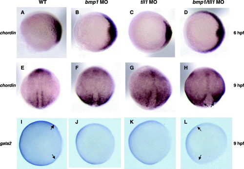
Altered expression patterns of chordin and gata2 in embryos dorsalized by knockdown of bmp1 and tll1. (A–D) Chordin localization is shown at 6 hpf for (A) wild type embryo; (B) bmp1 morphant, in which there is a lateral expansion of chordin expression; (C) tll1 morphant, which does not have a clearly expanded chordin expression domain; and (D) bmp1/tll1 double morphant, showing massive lateral expansion of chordin expression. (E–H) Chordin localization is shown at 9 hpf for (E) wild type embryos; (F) bmp1 morphant, in which chordin expression is expanded; (G) tll1 morphant, showing expanded chordin expression; and (H) bmp1/tll1 double morphant. The notochord is delineated by strong chordin signal in E–H, and by arrows in H. Note the broadening of the notochord observed in the dorsalized morphants. (I–L) gata2 localization is shown at 9 hpf. Note the greatly reduced gata2 expression domain in the bmp1/tll1 double morphant compared to wild type (gata2 domain margins marked by arrows in I and L). (A–D, I–L) Animal pole views, dorsal to the right. (E–H) Dorsal views, G and H slightly tilted to the right. EXPRESSION / LABELING:
|
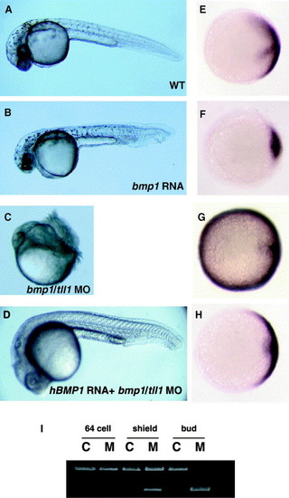
Specific role for BMP1 in zebrafish dorsoventral patterning. (A and B) Embryos either uninjected (A) or injected (B) with bmp1 RNA are shown at 55 hpf. (C and D) Embryos injected with both bmp1 MO1 and tll1 MO in the absence (C) or presence (D) of human BMP1 RNA are shown at 24 hpf. (E–H) Embryos in which in situ hybridization for chordin has been performed are shown at 75% epiboly, to the right of embryos injected with the same RNAs and/or MOs. Animal views are shown for E–H, with dorsal to the right. (I) RT-PCR analysis is shown for control uninjected embryos (C) or embryos injected with bmp1 SP-MO (M). EXPRESSION / LABELING:
|
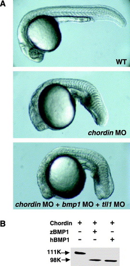
Chordin function is epistatic to bmp1 and Chordin is cleaved by zebrafish BMP1 proteinase. (A) Embryos uninjected (WT) or injected with chordin MO, or with a combination of chordin, bmp1, and tll1 MOs are shown at 24 hpf. (B) A Western blot is shown of murine Chordin incubated in the absence (-) or presence (+) of zebrafish (zBMP1) or human (hBMP1) BMP1 protein. The anti-Chordin antibody and Western blot conditions have been previously described (Pappano et al., 2003). |

Unillustrated author statements EXPRESSION / LABELING:
|
Reprinted from Mechanisms of Development, 123(7), Jasuja, R., Voss, N., Ge, G., Hoffman, G.G., Lyman-Gingerich, J., Pelegri, F., and Greenspan, D.S., bmp1 and mini fin are functionally redundant in regulating formation of the zebrafish dorsoventral axis, 548-558, Copyright (2006) with permission from Elsevier. Full text @ Mech. Dev.