- Title
-
Moz-dependent Hox expression controls segment-specific fate maps of skeletal precursors in the face
- Authors
- Crump, J.G., Swartz, M.E., Eberhart, J.K., and Kimmel, C.B.
- Source
- Full text @ Development
|
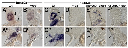
Moz is required for Hox expression in multiple facial tissues. Horizontal sections were taken at 34 hpf at dorsal (A'-F') and ventral (A"-F") levels of the pharyngeal segments and stained with probes against hoxb2a (A,B) or hoxa2b (C-F). The second and third segments (numbered) from individual sides are shown with anterior to the top and lateral (ectoderm) to the left. (A',A") In the wild-type pharynx, hoxb2a is expressed exclusively in CNC of the second segment. (B',B") hoxb2a expression is very reduced inmoz mutants. (C',C") In wild types, hoxa2b is strongly expressed in the CNC of the second and more posterior segments but not in the mesoderm core of the second segment. Ventral sections show additional expression in a small amount of surface ectoderm (arrows). In other sections, hoxa2b expression is seen in the endoderm of the second and more posterior pouches (data not shown). (D',D") In moz mutants, hoxa2b expression is absent or very reduced in CNC, surface ectoderm and endoderm. (E',E") In b1092 mutant sides that received moz- CNC transplants (see text), hoxa2b expression is lost in CNC. In this example, a small amount of CNC expression persists (arrowheads). However, weak expression of hoxa2b in the ectoderm is still seen (arrow). (F',F") In moz- sides that received wild-type ectoderm transplants (see text), hoxa2b expression is present in the ectoderm (arrow) but absent in CNC. EXPRESSION / LABELING:
|
|
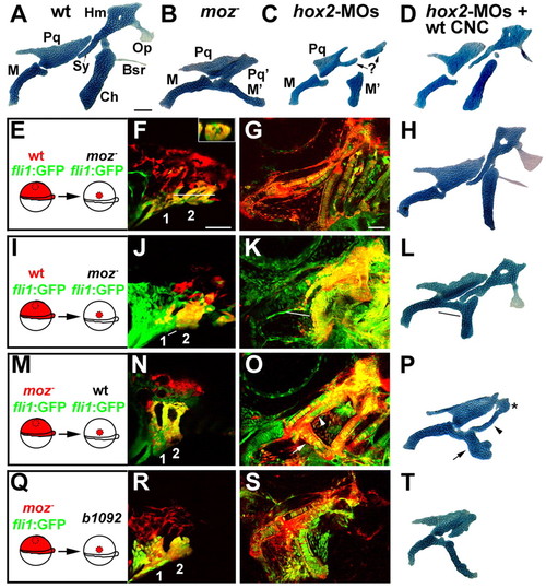
Moz and Hox2 genes function in CNC to control second-segment skeletal identity. (A-D,H,L,P,T) Facial skeletons from 4 dpf larvae. (A) Wild type. (B) moz- control side of H. (C) hox2-MO animal (control side of D). (D) hox2-MO animal unilaterally rescued with wild-type CNC. (E,I) Schematics showing unilateral transplantation of wild-type CNC precursors (red) into moz- hosts. fli1:GFP labels all CNC green. Wild-type CNC (red) populate the second segment (2) at 32 hpf (F,J) and by 4 dpf (G,H,K,L) form largely normal Hm, Sy, Ch, Op and Br skeletal elements in 8/10 moz- hosts. Inset in F is a digital longitudinal section through the white line and shows co-localization of the transplant lineage tracer and the fli1:GFP. Lateral is up. In I-L, a hybrid skeleton was observed. In this example, M'-like (black line in L) and Ch cartilages were fused together, with M' consisting of moz- host CNC (white lines in J and K), and Hm, Sy and Ch cartilages and Op osteocytes consisting entirely of wild-type donor CNC. (M) Schematic of transplantation of moz- CNC (red and green) into wild-type hosts. Both donor and host are fli1:GFP+. (N) moz- CNC (red) populate the second segment (2) at 36 hpf. (O,P) At 4 dpf, partial PQ'-like (*) and M'-like (arrow) cartilages consist of moz- donor CNC (white arrow), and a Sy-like element (arrowheads) forms primarily from wild-type host CNC. In 9/10 cases, moz- CNC precursors formed jaw-like cartilages in a wild-type host. (Q) Schematic of transplantation of moz-; fli1:GFP CNC (red and green) into b1092 hosts (note that hosts are fli1:GFP-). moz- CNC populate the first two segments (1,2) at 32 hpf (R) and by 4 dpf form moz--like M' and Pq' cartilages (S,T) in 6/9 moz+ hosts. Scale bars: 50 µm. |
|
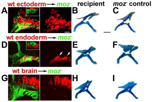
Moz is not sufficient in the endoderm, surface ectoderm or hindbrain to rescue moz- skeletal development. Wild-type ectoderm (A-C), endoderm (D-F) and hindbrain (G-I) precursors from fli1:GFP animals were unilaterally transplanted into moz-; fli1:GFP hosts at shield stage. Confocal images show that transplants (red) contributed significantly to the surface ectoderm (A), facial endoderm and pouches (arrows in D), and hindbrain (G) at 36 hpf. Insets in A and D are digital longitudinal sections through the level of the white lines and show that transplanted tissue did not express the fli1:GFP CNC marker. Lateral is up. (B,C,E,F,H,I) Facial skeletons of the first two segments from the transplanted animals at 4 dpf. Cartilages from the sides receiving transplants (B,E,H) were indistinguishable from the contralateral control sides (C,F,I). No rescue was seen in 3 ectoderm, 9 endoderm and 12 brain transplants. Scale bars: 50 µm. |
|
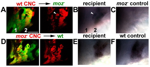
Moz controls hoxb2a expression cell-autonomously in CNC.(A,D) Confocal images at 36 hpf of the first two segments (numbered in A) of fli1:GFP hosts after unilateral transplantation of CNC precursors (red) at shield stage. After live imaging, the same animals were fixed and stained with hoxb2a RNA probe. The transplant recipient sides (B,E) and the contralateral control sides that received no transplant (C,F) are shown. (A-C) Wild-type CNC in a moz-; fli1:GFP host cell-autonomously rescue hoxb2a expression in the second segment. Note the similar zones of red donor and hoxb2a-expressing CNC in A and B; white arrowheads denote a small group of isolated CNC rescued for hoxb2a expression. Second segments of control moz- sides do not express hoxb2a (C). (D-F) moz- CNC in a wild-type fli1:GFP host cell-autonomously fail to express hoxb2a in the second segment. Note the reciprocal zones of red donor and hoxb2a-expressing CNC in D and E. Second segments of control wild-type sides express hoxb2a (F). The similarity of hoxb2a expression in B and E shows that non-hoxb2a-expressing cells preferentially sort to the anterior ventral domain of the second segment. Scale bar: 50 µm. EXPRESSION / LABELING:
|
|
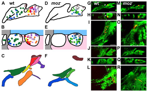
The fate map of second-segment skeletal derivatives is altered in moz mutants. Summaries of wild-type (A-C) and moz- (D-F) fatemaps and representative examples (G-R). Lateral views (A,D,G,J,M,P) and dorsal cross-sectional views at the level of the labelled cell with anterior to the left and lateral up (B,E,H,K,N,Q) are shown at 24 hpf. The facial skeleton is shown at 4 dpf (C,F,I,L,O,R). Filled circles denote facial cartilage precursors, and open circles denote precursors of neurocranial cartilage that surrounds the otic capsule. Triangles and squares denote precursors of the Op (upper) and Br (lower) dermal bones, respectively. moz- CNC that made no skeleton are hatched squares. In B and E, areas of surface ectoderm (blue), endoderm (pink), and stomodeal ectoderm (grey) are shown (compare to insets in Fig. 3A,D). In A,B,G,H, first pouch endoderm (P) is labelled. Wild-type examples of anterior Hm cartilage (G-I) and Op osteocyte (J-L) precursors are shown. (M-R) moz- CNC in similar positions fail to make skeleton. Note that in both wild types and moz mutants the most dorsal CNC in the first and second segment contribute to neurocranial cartilage that surrounds the otic capsule. Scale bars: 50 µm. |
|
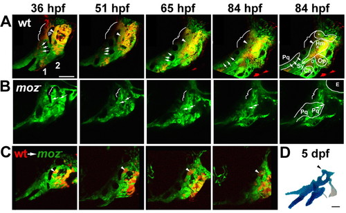
Moz is required for the growth and chondrification of dorsal second-segment CNC. (A-C) Progressive frames, as indicated, from time-lapse confocal recordings of first (1) and second (2) pharyngeal segment development in wild-type fli1:GFP (A), moz-; fli1:GFP (B), and moz-; fli1:GFP/wild-type fli1:GFP mosaic (C) animals. Skeletal outlines are shown at 84 hpf. (A) A subset of CNC precursors are labelled with a red dye. In wild type (n=4), dorsal second-segment CNC (arrowheads) adjacent to p1 endoderm (P) form the anterior part of Hm cartilage. Dorsal first-segment CNC (brackets) form either no cartilage or neurocranial cartilage (*). Intermediate first-segment CNC (arrows) contribute to Pq cartilage. (B) In moz-; fli1:GFP animals (n=6), intermediate second-segment CNC contribute to Pq' cartilage (arrows). More dorsal CNC make no cartilage (brackets). In C, small amounts of wild-type CNC precursors (red) were transplanted into moz-; fli1:GFP animals. Wild-type second-segment CNC (arrowheads) adjacent to p1 grow and form a cartilage nodule in the normally cartilage-free dorsal zone of the moz- host. (D) The skeletal preparation of this same animal at 5 dpf. Scale bars: 50 µm. See also Movies 1-3 in the supplementary material. |
|
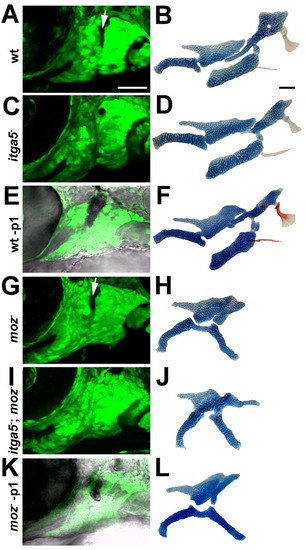
The development of moz- second-segment cartilage does not depend on p1 endoderm. CNC segments at 36 hpf (A,C,E,G,I,K) and facial skeletons at 4 dpf (B,D,F,H,J,L) from wild-type fli1:GFP (A,B,E,F), itga5b926; fli1:GFP (C,D), moz-; fli1:GFP (G,H,K,L), and moz-; itga5b926; fli1:GFP (I,J) animals. In E and K, inclusion of the Normarski channel shows that p1 endoderm has been selectively ablated by laser irradiation, as evinced by the dark pyknotic nuclei. When p1 is eliminated in either itga5b926; fli1:GFP (C) or p1 ablated (-p1) (E) animals, the anterior portion of the Hm cartilage is lost and Sy cartilage is variably reduced (D,F). However, removal of p1 in moz-; itga5b926; fli1:GFP (I) or p1 ablated moz- (K) animals does not affect the shape of Pq' (J,L). For p1 laser ablations, 13/14 wild-type animals had reduced Hm cartilage and 0/5 moz mutants had reduced Pq'. Arrows denote p1. Scale bars: 50 µm. |

