
- Title
-
Zebrafish furin mutants reveal intricacies in regulating Endothelin1 signaling in craniofacial patterning
- Authors
- Walker, M.B., Miller, C.T., Talbot, J.C., Stock, D.W., and Kimmel, C.B.
- Source
- Full text @ Dev. Biol.
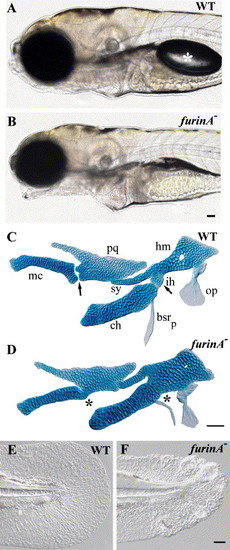
furinA mutants have jaw and fin defects. (A and B) Live views at 5 dpf. furinA mutants have an open-mouth phenotype and fail to form a swim bladder (white asterisk in panel A). (C and D) Flat mounts of Alcian green-stained cartilages at 5 dpf. Wild-type DV joint regions are indicated with arrows in panel C. Fusions at joint regions in furinA mutants are indicated with asterisks in panel D. Cartilages are labeled as followed: pq (palatoquadrate), mc (Meckel's cartilage), hm (hyomandibula), ch (ceratohyal), sy (symplectic), and ih (interhyal). Two bones of the hyoid arch are also lightly stained with Alcian green: op (opercle), bsrp (branchiostegal ray posterior). (E and F) Live views of the tail fin at 2 dpf. furinA mutant larvae have mildly ruffled fins. Scale bars: 50 μm. |
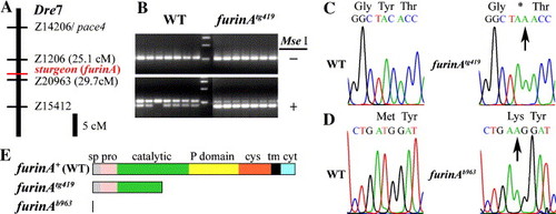
The sturgeon gene is furinA. (A) sturgeon (furinA) maps to Danio rerio chromosome 7. (B) PCR genotyping illustrating co-segregation of nonsense lesion with furinAtg419 mutant phenotype following MseI digestion. (C and D) Sequence chromatograms of furinA+ (wt), furinAtg419, and furinAb963 alleles around lesion sites in furinA. Arrows indicate lesions. (E) Schematic of protein domains in furinA+ (wt), furinAtg419, and furinAb963 alleles. Abbreviations: sp (signal peptide), pro (propeptide), cys (cysteine rich), tm (transmembrane), cyt (cytosolic). |
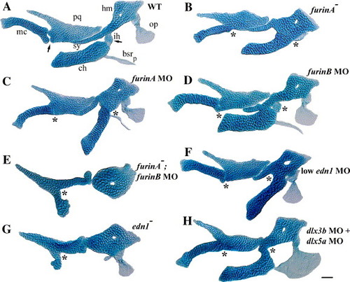
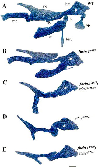
Both FurinA and FurinB function are required to activate Edn1 signaling. Flat mounts of Alcian-stained cartilages and bones at 5–6 dpf. (A) Wild-type, (B) furinA mutant, (C) furinA MO-injected larva, (D) furinB MO-injected larva, (E) furinB MO injected into furinA mutant, (F) low edn1 MO-injected larva, (G) edn1 mutant, and (H) dlx3b MO + dlx5a MO-injected larva. Wild-type DV joint regions are indicated with arrows in panel A. Fusions at joint regions are indicated with asterisks in panels B–H. Scale bar: 50 μm. PHENOTYPE:
|
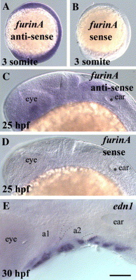
furinA expression overlaps with edn1 expression. furinA antisense and sense in situ hybridizations at 3 somite stage (11 hpf) (A and B) and 25 hpf (C and D). (E) edn1 in situ hybridization at 30 hpf. a1 and a2 label pharyngeal arch 1 and 2. Dotted lines label pharyngeal pouches 1 and 2. Scale bar: 50 μm. EXPRESSION / LABELING:
|
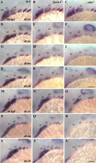
furinA is required for early but not late edn1-dependent Dlx gene expression. Dlx expression in wild-type, furinA mutant, and edn1 mutant embryos at 30 hpf (A–L) and 36 hpf (M–U). (A–C) dlx2a, (D–F, M–O) dlx5a, (G–I, P–R) dlx6a, and (J–L, S–U) dlx3b. dlx2a expression is slightly affected in both furinA and edn1 mutant embryos, whereas expression of dlx3b, dlx5a, and dlx6a is moderately reduced in furinA mutants at 30 hpf and strongly reduced in edn1 mutants. Expression of dlx5a, dlx6a, and dlx3b recovers significantly in furinA mutants by 36 hpf. a1 and a2 label pharyngeal arch 1 and 2. Dotted lines label pharyngeal pouches 1 and 2 in (AU) and bottom of arch 1 and 2 in (O, R, and U). Scale bars: 50 μm. |
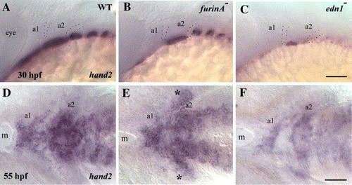
furinA is required for early but not late edn1-dependent hand2 expression. Lateral views of hand2 expression in wild-type (A), furinA mutant (B), and edn1 mutant (C) embryos at 30 hpf. Ventral views at 55 hpf (D–F). At 30 hpf, hand2 expression in furinA is strongly reduced in the first arch and moderately reduced in the second arch but recovers in both arches by 55 hpf. hand2 expression in edn1 mutants is strongly reduced in both arches at 30 and 55 hpf. a1 and a2 label pharyngeal arch 1 and 2. Dotted lines label pharyngeal pouches 1 and 2. Asterisks in panel E mark ectopic expression of hand2 in the second arch in furinA mutants at 55 hpf. m, mouth. Scale bars: 50 μm. |
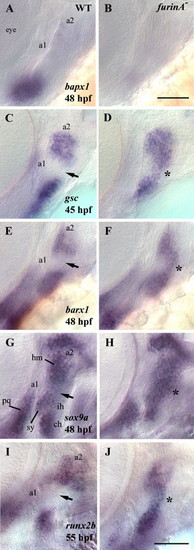
Intermediate arch domains are misspecified in furinA mutants. (A, B) bapx1 expression is strongly reduced in furinA mutants at 48 hpf. (C, D) At 45 hpf, gsc is ectopically expressed in a second arch intermediate domain in furinA mutants. (E, F) barx1 is ectopically expressed in a second arch intermediate domain in furinA mutants at 48 hpf. (G, H) sox9a is ectopically expressed in a second arch intermediate domain in furinA mutants at 48 hpf. (I, J) runx2b is ectopically expressed in a second arch intermediate domain in furinA mutants at 55 hpf. Arrows indicate a second arch intermediate domain in wild-type embryos. Asterisks indicate ectopic expression in a second arch intermediate domain in furinA mutant embryos. a1 and a2 label pharyngeal arch 1 and 2. Scale bars: 50 μm. |
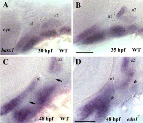
barx1 is ectopically expressed in intermediate arch domain in edn1 mutants. barx1 expression in wild-type embryos at 30 hpf (A), 35 hpf (B), and 48 hpf (C), and in edn1 mutant embryos at 48 hpf (D). barx1 expression is ectopically expressed in the intermediate arch domains of edn1 mutants, and reduced in a second arch ventral domain at 48 hpf. Arrows indicate intermediate arch domains in wild-type embryos. Asterisks indicate ectopic expression in intermediate arch domain in edn1 mutant embryos. a1 and a2 label pharyngeal arch 1 and 2. Dotted lines label pharyngeal pouches 1 and 2. Scale bars: 50 μm. |
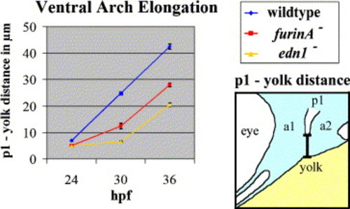
furinA and edn1 mutants have an early defect in ventral arch elongation. Bottom of pharyngeal pouch 1 (p1) to yolk distance was measured in micrometers. Each data point represents measurements from 5 embryos, with error bars representing standard error. edn1 mutants have a strong defect in ventral arch elongation from 24 to 30 hpf. furinA mutants have a moderate defect in ventral arch elongation from 24 to 30 hpf. The ventral arch elongation defects recover significantly in furinA and edn1 mutants between 30 and 36 hpf. |
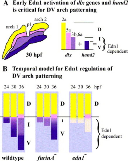
Edn1 signaling specifies arch fates through regulation of Dlx gene expression and ventral arch elongation. (A) Edn1 activation of Dlx genes and hand2 is critical for DV pharyngeal arch patterning. Edn1 activation of hand2 subdivides a broader dlx3b and dlx6a domain into intermediate and ventral arch fates. Dorsal arch fates are largely Edn1 independent. (B) Temporal model for Edn1 regulation of intermediate/ventral (I/V) domain patterning. Early Edn1 signaling specifies an arch compartment, containing both intermediate and ventral fates. Subsequent segregation of cell fates occurs in conjunction with ventral arch elongation. furinA and edn1 mutants have a delay in ventral arch elongation, and defects in the specification and segregation of cell fates. Black line represents ventral aspect of first pharyngeal pouch. Bars below black line represent ventral arch and are drawn to scale with ventral arch measurements from Fig. 10. Colors represent response of cells to Edn1 signaling as in panel A. Yellow bars above black line represent dorsal arch and are not drawn to scale. (D) dorsal, (I) intermediate, (V) ventral, (p1) pharyngeal pouch 1. |
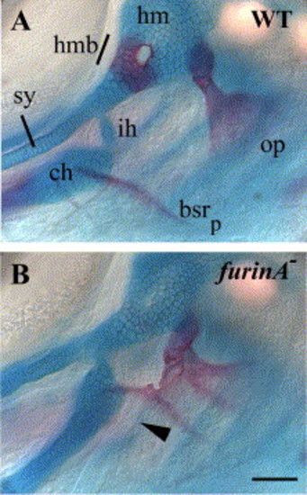
furinA mutants have bony fusions and altered muscle insertions points near joint domains. (A and B) Whole mounts of WT and furinA mutant larvae at 6 dpf doubly stained with Alcian blue and Alizarin red for cartilage and bone, respectively. furinA mutants have a low penetrance of fusions between opercle (op) and posterior branchiostegal ray (bsrp) bones. Hmb refers to hyomandibular chondral bone. Arrowhead points to altered muscle near joint region in furinA mutants. Scale bars: 50 um. |

Loss of maternal furinA does not enhance jaw defects in furinA mutants. (A) Temporal expression profiles of furinA and edn1 were determined by RT-PCR. The zebrafish ornithinedecarboxylase (odc) gene was used as an internal PCR control. The following primer pairs were used: furinA, 5′AGCGGAGGCGTCAATGA3′/5′TGAGCACGGCCACAATGGCAG3′ (1138 bp); edn1, 5′CCTGAAATGCATGACGTGTG3′/5′AATACGGGACTTGCATACTACA3′ (659 bp); and odc, 52ACACTATGACGGCTTGCACCG3′/5′CCCACTGACTGCACGATCTGG3′ (309 bp). (B) Loss of maternal furinA alone yields only phenotypically wild-type larvae. Loss of both maternal and zygotic furinA does not enhance joint loss phenotype over loss of zygotic furinA alone. M, maternal; Z, zygotic. |
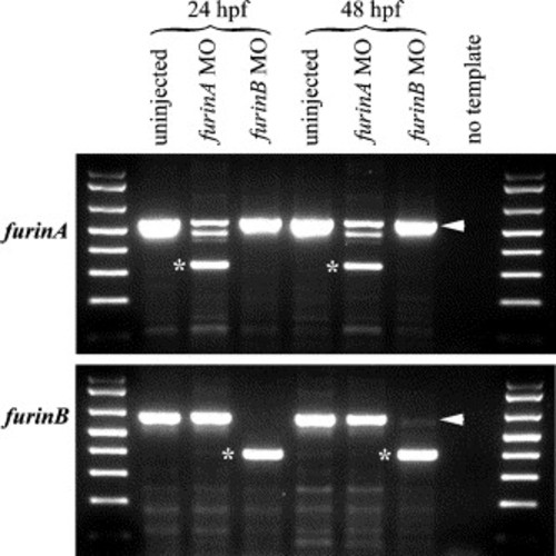
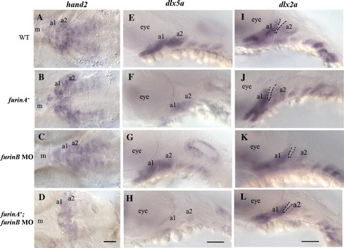
furinA and furinB redundantly activate dlx5a, hand2, and stimulate ventral arch elongation. (A–D) hand2 expression at 55 hpf is dramatically reduced in furinA−;furinB MO (D) embryos with respect to WT (A), furinA− (B), and furinB MO (C) embryos. (E–H) dlx5a expression at 36 hpf is strongly reduced in furinA−;furinB MO (H) embryos with respect to WT (E), furinA− (F), and furinB MO (G) embryos. (I–L) dlx2a staining at 36 hpf illustrates the arch length. Ventral arch length below the first pouch is substantially reduced in furinA−;furinB MO embryos (L) with respect to WT (I), furinA− (J), and furinB MO (K) embryos. a1 and a2 label pharyngeal arch 1 and 2. Dotted lines label pouch 1. m labels the mouth; eye labels the eye. Scale bars: 50 μm. |

Unillustrated author statements PHENOTYPE:
|
Reprinted from Developmental Biology, 295(1), Walker, M.B., Miller, C.T., Talbot, J.C., Stock, D.W., and Kimmel, C.B., Zebrafish furin mutants reveal intricacies in regulating Endothelin1 signaling in craniofacial patterning, 194-205, Copyright (2006) with permission from Elsevier. Full text @ Dev. Biol.