
- Title
-
fused-somites-like mutants exhibit defects in trunk vessel patterning
- Authors
- Shaw, K.M., Castranova, D.A., Pham, V.N., Kamei, M., Kidd, K.R., Lo, B.D., Torres-Vazquez, J., Ruby, A., and Weinstein, B.M.
- Source
- Full text @ Dev. Dyn.
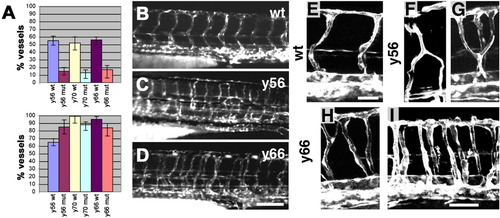
Intersegmental vessel migration and patterning are defective in y56/y70 and y55/y66 mutants. A: Quantitation of the percent of intersegmental vessel sprouts present in y56, y70, and y66 mutants and their phenotypically wild-type siblings at 22 hours postfertilization (hpf; top), and the number of intersegmental vessel sprouts that have reached or passed the horizontal myoseptum at 30 hpf (bottom). B-D: Fluorescence images of trunk/tail vessels in 48 hpf TG(fli1:egfp)y1 wild-type (B), y56 mutant (C), and y66 mutant (D) animals. Note the regular alternating pattern of vessels in wild-types compared with the irregular arrangement in mutants. E-I: Higher magnification confocal images of trunk vessels in 48 hpf TG(fli1:egfp)y1 wild-type (E), y56 mutant (F,G), and y66 mutant (H,I) animals. Intersegmental vessels in mutant animals display dual ventral roots, ectopic dorsal branching, and lack of left-right mirroring of vessel segments. Images in B-I are all oriented with anterior to the left and dorsal up. Scale bars = 100 μm in B-D, 25 μm in E,F, 50 μm in G-I. PHENOTYPE:
|
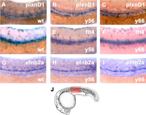
Endothelial markers are appropriately expressed in y56/y70 and y55/y66 mutants. A-I: Whole-mount in situ hybridization of mid-trunk vessels in 24 hours postfertilization (hpf) wild-type (A,D,G), y56 mutant (B,E,H), and y66 mutant (C,F,I) embryos using probes for plxnD1 (A-C), flt4 (D-F), and efnb2a (G-I). J: The region shown in the images in A-I is highlighted in red. Endothelial markers are appropriately expressed in mutants, with restriction of efnb2a and flt4 to arterial and venous vessels, respectively. Anterior is to the left and dorsal is up in all panels. The camera lucida drawing in J is modified from Kimmel et al. (1995). EXPRESSION / LABELING:
|
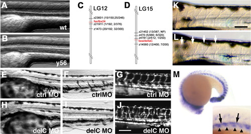
Characterization of somite patterning defects and molecular analysis of y56/y70 and y55/y66 mutants. A,B: Transmitted light micrographs of the trunks of 72 hours postfertilization (hpf) wild-type (A) and y56 mutant (B) animals, showing poor morphogenesis of somite boundaries in mutants. C,D: Genetic mapping of y56 and y70 (C) and y55 and y66 (D) mutants. C: y56 and y70 mutants are tightly linked to tbx24 on LG12. The numbers in parentheses show linkage (number of recombinants/total meioses) to CA repeat markers for y56 (first numbers in parentheses) and y70 (last numbers in parentheses). D: The y55 and y66 mutants are tightly linked to deltaC on LG15. The numbers in parentheses show linkage (number of recombinants/total meioses) to CA repeat markers for y55 (first numbers in parentheses) and y66 (last numbers in parentheses). NP indicates marker was not polymorphic for particular mutant and marker combination. E-L: Transmitted light (E,F,H,I) and TG(fli1:egfp)y1 vascular fluorescence (G,J) micrographs of 48 hpf trunks (E,G,H,J) and tails (F,I) of control (E-G) and deltaC (H-J) morpholino injected animals. H,I: Animals injected with deltaC morpholinos form somite boundaries 1-4 (H) but not more posterior boundaries (I) properly and show defects in vessel patterning indistinguishable from those seen in y55 and y56 mutants (J). K,L: Higher magnification transmitted light images of the anterior trunk of 3 days postfertilization wild-type (K) and y56 mutant (L) animals, with well-formed anterior somites in y56 (arrows). Whole-mount in situ hybridization of a 19-20 hpf wild-type animal using a probe for delC. Magnified inset shows expression in dorsal aorta (arrows) and somites (arrowheads). Anterior is to the left and dorsal is up in all micrographs. Scale bars = 100 μm. PHENOTYPE:
|
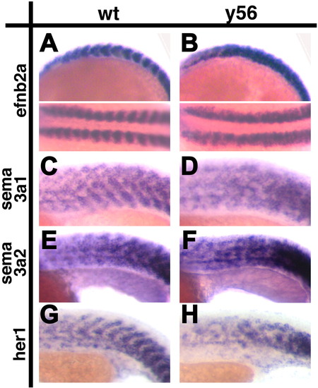
Disruption of somitic markers in y56 mutants. A-H: Whole-mount in situ hybridization of the trunk region of wild-type (A,C,E,G) and y56 mutant (B,D,F,H) animals at 16.5 hours postfertilization (hpf; A,B) and 19-21 hpf (C-H) using probes for efnb2a (A,B), sema3a1 (C,D), sema3a2 (E,F), and her1 (G,H). Somitic markers show a more disorganized pattern of expression in y56 mutants. Anterior is to the left and dorsal is up in all panels. EXPRESSION / LABELING:
|